Cutaneous Presentation of Metastatic Salivary Duct Carcinoma

Article Type
Changed
Tue, 10/17/2023 - 12:43
Display Headline
Cutaneous Presentation of Metastatic Salivary Duct Carcinoma

To the Editor:

Metastatic spread of salivary duct carcinoma (SDC) to the skin is rare. Diagnosing SDC can be challenging because the cutaneous manifestations of this disease are variable and include nodules, papules, and erysipelaslike inflammation (also known as shield sign) with purpuric papules and pseudovesicles. We describe a case of cutaneous metastatic SDC that originated from the parotid gland and presented with 2 distinct cutaneous findings: sharply demarcated erythematous plaques and focally hemorrhagic angiomatous papules.

A 60-year-old man presented with a persistent polymorphous pruritic eruption of several months’ duration involving the entire face, ears, neck, and upper chest. He had a history of unspecified adenocarcinoma of the parotid gland diagnosed 2 years prior and underwent multiple treatment cycles with several chemotherapeutic agents over the course of 18 months. Physical examination showed erythematous papules and nodules on the face and neck with slight overlying scale. Sharply demarcated, erythematous plaques studded with focally hemorrhagic, angiomatous papules were noted on the neck and chest (Figure 1). Two 4-mm punch biopsies were sampled from representative nodular areas. Histopathology showed multiple round solid-tumor nodules with central necrosis in the superficial and deep dermis that were not associated with the overlying epidermis (Figures 2A and 2B). The tumor cells appeared polygonal and contained ample eosinophilic cytoplasm. Tumor nuclei showed marked pleomorphism, and numerous atypical mitotic figures were readily identifiable (Figure 2C). There was diffuse cytoplasmic staining with cytokeratin 7 and nuclear staining with androgen receptor (Figure 2D). These findings were consistent with a diagnosis of SDC metastatic to the skin.

CT112004013_e_Fig_1_AB.jpg
%3Cp%3E%3Cstrong%3EFIGURE%201.%3C%2Fstrong%3E%20A%20and%20B%2C%20Sharply%20demarcated%2C%20erythematous%20plaques%20studded%20with%20focally%20hemorrhagic%2C%20angiomatous%20papules%20on%20the%20neck%20and%20upper%20chest%20in%20a%20patient%20with%20cutaneous%20metastatic%20salivary%20duct%20carcinoma%20that%20originated%20from%20the%20parotid%20gland.%3C%2Fp%3E

The patient underwent 8 cycles of docetaxel chemotherapy. With disease progression, the chemotherapy regimen was changed to gemcitabine and methotrexate. The patient continued to experience disease progression and died 9 months after diagnosis of skin metastases.

CT112004013_e_Fig2_ABCD.jpg
%3Cp%3E%3Cstrong%3EFIGURE%202.%3C%2Fstrong%3E%20A%2C%20Histopathology%20showed%20multiple%20round%20solid-tumor%20nodules%20in%20the%20superficial%20and%20deep%20dermis%20with%20massive%20comedo%20necrosis%20(H%26amp%3BE%2C%20original%20magnification%20%C3%9710).%20B%2C%20A%20tumor%20nodule%20with%20central%20zone%20of%20cellular%20necrosis%20(H%26amp%3BE%2C%20original%20magnification%20%C3%9720).%20C%2C%20The%20nuclei%20of%20tumor%20cells%20showed%20marked%20pleomorphism%20and%20atypical%20mitotic%20figures%20(H%26amp%3BE%2C%20original%20magnification%20%C3%9740).%20D%2C%20Immunohistochemistry%20expressed%20diffuse%20nuclear%20staining%20of%20tumor%20cells%20with%20androgen%20receptor%20(original%20magnification%20%C3%9720).%3C%2Fp%3E

Salivary duct carcinoma is rare and is estimated to represent 1% to 3% of all salivary malignancies.1 It is a highly aggressive form of salivary gland carcinoma and is associated with a poor clinical outcome. The 3-year overall survival rate for stage I disease is 42% and only 23% for stage IV disease.2 Salivary duct carcinoma has a high rate of distant metastasis,3 but cases of cutaneous metastases are rare.3-8 Previously reported cases of SDC that metastasized to the skin originated from the parotid gland (n=6) and submandibular gland (n=1).3

The diagnosis of cutaneous metastases is challenging due to the variability of the skin manifestations. Three cases described small firm nodules in patients,3-5 while others presented with purpuric papules and pseudovesicles.6-8 Our patient presented with sharply demarcated, erythematous plaques studded with focally hemorrhagic, angiomatous papules, which further emphasizes the capricious nature of skin findings.

The morphology of SDC is strikingly similar to ductal adenocarcinoma of the breast, which can lead to diagnostic confusion. Both carcinomas may show oncocytic cells, ductal formations, and cribriform structures with central comedo necrosis. Moreover, immunohistochemical features overlap, including positive staining for cytokeratin 7 and gross cystic disease fluid protein 15. Positive immunohistochemistry with androgen receptor is consistent with SDC but also can be expressed in some cases of breast carcinoma.9,10 Therefore, the diagnosis of cutaneous involvement from metastatic SDC requires not just an evaluation of the pathologic features but careful attention to the clinical history and a thorough staging evaluation.

References
  1. D’heygere E, Meulemans J, Vander Poorten V. Salivary duct carcinoma. Curr Opin Otolaryngol Head Neck Surg. 2018;26:142-151.
  2. Gilbert MR, Sharma A, Schmitt NC, et al. A 20-year review of 75 cases of salivary duct carcinoma. JAMA Otolaryngol Head Neck Surg. 2016;142:489-495. 
  3. Chakari W, Andersen L, Andersen JL. Cutaneous metastases from salivary duct carcinoma of the submandibular gland. Case Rep Dermatol. 2017;9:254-258.
  4. Tok J, Kao GF, Berberian BJ, et al. Cutaneous metastasis from a parotid adenocarcinoma. Report of a case with immunohistochemical findings and review of the literature. Am J Dermatopathol. 1995;17:303-306.
  5. Aygit AC, Top H, Cakir B, et al. Salivary duct carcinoma of the parotid gland metastasizing to the skin: a case report and review of the literature. Am J Dermatopathol. 2005;27:48-50.
  6. Cohen PR, Prieto VG, Piha-Paul SA, et al. The “shield sign” in two men with metastatic salivary duct carcinoma to the skin: cutaneous metastases presenting as carcinoma hemorrhagiectoides. J Clin Aesthet Dermatol. 2012;5:27-36.
  7. Hafiji J, Rytina E, Jani P, et al. A rare cutaneous presentation of metastatic parotid adenocarcinoma. Australas J Dermatol. 2013;54:E40-E42.
  8. Zanca A, Ferracini U, Bertazzoni MG. Telangiectatic metastasis from ductal carcinoma of the parotid gland. J Am Acad Dermatol. 1993;28:113-114.
  9. Brys´ M, Wójcik M, Romanowicz-Makowska H, et al. Androgen receptor status in female breast cancer: RT-PCR and Western blot studies. J Cancer Res Clin Oncol. 2002;128:85-90. 
  10. Udager AM, Chiosea SI. Salivary duct carcinoma: an update on morphologic mimics and diagnostic use of androgen receptor immunohistochemistry. Head Neck Pathol. 2017;11:288-294.
Article PDF
Author and Disclosure Information

Drs. Wang, Vyas, Alghamdi, Sapadin, Goldberg, and Westra are from the Icahn School of Medicine, Mount Sinai, New York, New York. Drs. Wang and Westra are from the Department of Pathology, Drs. Vyas and Goldberg are from the Departments of Pathology and Dermatology, and Drs. Alghamdi and Sapadin are from the Department of Dermatology. Dr. Parker is from the Albert Einstein College of Medicine at Mount Sinai, Bronx, New York.

The authors report no conflict of interest.

Correspondence: William Westra, MD, Department of Pathology, Icahn School of Medicine at Mount Sinai, 1468 Madison Ave, New York, NY 10029 (william.westra@mountsinai.org).

Issue
Cutis - 112(4)
Publications
Topics
Page Number
E13-E15
Sections
Author and Disclosure Information

Drs. Wang, Vyas, Alghamdi, Sapadin, Goldberg, and Westra are from the Icahn School of Medicine, Mount Sinai, New York, New York. Drs. Wang and Westra are from the Department of Pathology, Drs. Vyas and Goldberg are from the Departments of Pathology and Dermatology, and Drs. Alghamdi and Sapadin are from the Department of Dermatology. Dr. Parker is from the Albert Einstein College of Medicine at Mount Sinai, Bronx, New York.

The authors report no conflict of interest.

Correspondence: William Westra, MD, Department of Pathology, Icahn School of Medicine at Mount Sinai, 1468 Madison Ave, New York, NY 10029 (william.westra@mountsinai.org).

Article PDF
Article PDF

To the Editor:

Metastatic spread of salivary duct carcinoma (SDC) to the skin is rare. Diagnosing SDC can be challenging because the cutaneous manifestations of this disease are variable and include nodules, papules, and erysipelaslike inflammation (also known as shield sign) with purpuric papules and pseudovesicles. We describe a case of cutaneous metastatic SDC that originated from the parotid gland and presented with 2 distinct cutaneous findings: sharply demarcated erythematous plaques and focally hemorrhagic angiomatous papules.

A 60-year-old man presented with a persistent polymorphous pruritic eruption of several months’ duration involving the entire face, ears, neck, and upper chest. He had a history of unspecified adenocarcinoma of the parotid gland diagnosed 2 years prior and underwent multiple treatment cycles with several chemotherapeutic agents over the course of 18 months. Physical examination showed erythematous papules and nodules on the face and neck with slight overlying scale. Sharply demarcated, erythematous plaques studded with focally hemorrhagic, angiomatous papules were noted on the neck and chest (Figure 1). Two 4-mm punch biopsies were sampled from representative nodular areas. Histopathology showed multiple round solid-tumor nodules with central necrosis in the superficial and deep dermis that were not associated with the overlying epidermis (Figures 2A and 2B). The tumor cells appeared polygonal and contained ample eosinophilic cytoplasm. Tumor nuclei showed marked pleomorphism, and numerous atypical mitotic figures were readily identifiable (Figure 2C). There was diffuse cytoplasmic staining with cytokeratin 7 and nuclear staining with androgen receptor (Figure 2D). These findings were consistent with a diagnosis of SDC metastatic to the skin.

CT112004013_e_Fig_1_AB.jpg
%3Cp%3E%3Cstrong%3EFIGURE%201.%3C%2Fstrong%3E%20A%20and%20B%2C%20Sharply%20demarcated%2C%20erythematous%20plaques%20studded%20with%20focally%20hemorrhagic%2C%20angiomatous%20papules%20on%20the%20neck%20and%20upper%20chest%20in%20a%20patient%20with%20cutaneous%20metastatic%20salivary%20duct%20carcinoma%20that%20originated%20from%20the%20parotid%20gland.%3C%2Fp%3E

The patient underwent 8 cycles of docetaxel chemotherapy. With disease progression, the chemotherapy regimen was changed to gemcitabine and methotrexate. The patient continued to experience disease progression and died 9 months after diagnosis of skin metastases.

CT112004013_e_Fig2_ABCD.jpg
%3Cp%3E%3Cstrong%3EFIGURE%202.%3C%2Fstrong%3E%20A%2C%20Histopathology%20showed%20multiple%20round%20solid-tumor%20nodules%20in%20the%20superficial%20and%20deep%20dermis%20with%20massive%20comedo%20necrosis%20(H%26amp%3BE%2C%20original%20magnification%20%C3%9710).%20B%2C%20A%20tumor%20nodule%20with%20central%20zone%20of%20cellular%20necrosis%20(H%26amp%3BE%2C%20original%20magnification%20%C3%9720).%20C%2C%20The%20nuclei%20of%20tumor%20cells%20showed%20marked%20pleomorphism%20and%20atypical%20mitotic%20figures%20(H%26amp%3BE%2C%20original%20magnification%20%C3%9740).%20D%2C%20Immunohistochemistry%20expressed%20diffuse%20nuclear%20staining%20of%20tumor%20cells%20with%20androgen%20receptor%20(original%20magnification%20%C3%9720).%3C%2Fp%3E

Salivary duct carcinoma is rare and is estimated to represent 1% to 3% of all salivary malignancies.1 It is a highly aggressive form of salivary gland carcinoma and is associated with a poor clinical outcome. The 3-year overall survival rate for stage I disease is 42% and only 23% for stage IV disease.2 Salivary duct carcinoma has a high rate of distant metastasis,3 but cases of cutaneous metastases are rare.3-8 Previously reported cases of SDC that metastasized to the skin originated from the parotid gland (n=6) and submandibular gland (n=1).3

The diagnosis of cutaneous metastases is challenging due to the variability of the skin manifestations. Three cases described small firm nodules in patients,3-5 while others presented with purpuric papules and pseudovesicles.6-8 Our patient presented with sharply demarcated, erythematous plaques studded with focally hemorrhagic, angiomatous papules, which further emphasizes the capricious nature of skin findings.

The morphology of SDC is strikingly similar to ductal adenocarcinoma of the breast, which can lead to diagnostic confusion. Both carcinomas may show oncocytic cells, ductal formations, and cribriform structures with central comedo necrosis. Moreover, immunohistochemical features overlap, including positive staining for cytokeratin 7 and gross cystic disease fluid protein 15. Positive immunohistochemistry with androgen receptor is consistent with SDC but also can be expressed in some cases of breast carcinoma.9,10 Therefore, the diagnosis of cutaneous involvement from metastatic SDC requires not just an evaluation of the pathologic features but careful attention to the clinical history and a thorough staging evaluation.

References
  1. D’heygere E, Meulemans J, Vander Poorten V. Salivary duct carcinoma. Curr Opin Otolaryngol Head Neck Surg. 2018;26:142-151.
  2. Gilbert MR, Sharma A, Schmitt NC, et al. A 20-year review of 75 cases of salivary duct carcinoma. JAMA Otolaryngol Head Neck Surg. 2016;142:489-495. 
  3. Chakari W, Andersen L, Andersen JL. Cutaneous metastases from salivary duct carcinoma of the submandibular gland. Case Rep Dermatol. 2017;9:254-258.
  4. Tok J, Kao GF, Berberian BJ, et al. Cutaneous metastasis from a parotid adenocarcinoma. Report of a case with immunohistochemical findings and review of the literature. Am J Dermatopathol. 1995;17:303-306.
  5. Aygit AC, Top H, Cakir B, et al. Salivary duct carcinoma of the parotid gland metastasizing to the skin: a case report and review of the literature. Am J Dermatopathol. 2005;27:48-50.
  6. Cohen PR, Prieto VG, Piha-Paul SA, et al. The “shield sign” in two men with metastatic salivary duct carcinoma to the skin: cutaneous metastases presenting as carcinoma hemorrhagiectoides. J Clin Aesthet Dermatol. 2012;5:27-36.
  7. Hafiji J, Rytina E, Jani P, et al. A rare cutaneous presentation of metastatic parotid adenocarcinoma. Australas J Dermatol. 2013;54:E40-E42.
  8. Zanca A, Ferracini U, Bertazzoni MG. Telangiectatic metastasis from ductal carcinoma of the parotid gland. J Am Acad Dermatol. 1993;28:113-114.
  9. Brys´ M, Wójcik M, Romanowicz-Makowska H, et al. Androgen receptor status in female breast cancer: RT-PCR and Western blot studies. J Cancer Res Clin Oncol. 2002;128:85-90. 
  10. Udager AM, Chiosea SI. Salivary duct carcinoma: an update on morphologic mimics and diagnostic use of androgen receptor immunohistochemistry. Head Neck Pathol. 2017;11:288-294.
Issue
Cutis - 112(4)
Issue
Cutis - 112(4)
Page Number
E13-E15
Page Number
E13-E15
Publications
Publications
Topics
Article Type
Display Headline
Cutaneous Presentation of Metastatic Salivary Duct Carcinoma
Display Headline
Cutaneous Presentation of Metastatic Salivary Duct Carcinoma
Sections
Teambase XML
<?xml version="1.0" encoding="UTF-8"?>
<!--$RCSfile: InCopy_agile.xsl,v $ $Revision: 1.35 $-->
<!--$RCSfile: drupal.xsl,v $ $Revision: 1.7 $-->
<root generator="drupal.xsl" gversion="1.7"> <header> <fileName>Wang 1023</fileName> <TBEID>0C02E6A4.SIG</TBEID> <TBUniqueIdentifier>NJ_0C02E6A4</TBUniqueIdentifier> <newsOrJournal>Journal</newsOrJournal> <publisherName>Frontline Medical Communications Inc.</publisherName> <storyname>Wang</storyname> <articleType>1</articleType> <TBLocation>Copyfitting-CT</TBLocation> <QCDate/> <firstPublished>20231017T111952</firstPublished> <LastPublished>20231017T111952</LastPublished> <pubStatus qcode="stat:"/> <embargoDate/> <killDate/> <CMSDate>20231017T111952</CMSDate> <articleSource/> <facebookInfo/> <meetingNumber/> <byline>Xintong Wang, MD; Nikki S. Vyas, MD; Abdulaziz A. Alghamdi, MD</byline> <bylineText>Xintong Wang, MD; Nikki S. Vyas, MD; Abdulaziz A. Alghamdi, MD; Matthew Parker, MD; Allen Sapadin, MD; Matthew S. Goldberg, MD; William Westra, MD </bylineText> <bylineFull>Xintong Wang, MD; Nikki S. Vyas, MD; Abdulaziz A. Alghamdi, MD</bylineFull> <bylineTitleText/> <USOrGlobal/> <wireDocType/> <newsDocType/> <journalDocType/> <linkLabel/> <pageRange>E13-E15</pageRange> <citation/> <quizID/> <indexIssueDate/> <itemClass qcode="ninat:text"/> <provider qcode="provider:"> <name/> <rightsInfo> <copyrightHolder> <name/> </copyrightHolder> <copyrightNotice/> </rightsInfo> </provider> <abstract/> <metaDescription>To the Editor:Metastatic spread of salivary duct carcinoma (SDC) to the skin is rare. Diagnosing SDC can be challenging because the cutaneous manifestations of </metaDescription> <articlePDF>298584</articlePDF> <teaserImage/> <title>Cutaneous Presentation of Metastatic Salivary Duct Carcinoma</title> <deck/> <disclaimer/> <AuthorList/> <articleURL/> <doi/> <pubMedID/> <publishXMLStatus/> <publishXMLVersion>1</publishXMLVersion> <useEISSN>0</useEISSN> <urgency/> <pubPubdateYear>2023</pubPubdateYear> <pubPubdateMonth>October</pubPubdateMonth> <pubPubdateDay/> <pubVolume>112</pubVolume> <pubNumber>4</pubNumber> <wireChannels/> <primaryCMSID/> <CMSIDs> <CMSID>2163</CMSID> </CMSIDs> <keywords> <keyword>nonmelanoma skin cancer</keyword> </keywords> <seeAlsos/> <publications_g> <publicationData> <publicationCode>CT</publicationCode> <pubIssueName>October 2023</pubIssueName> <pubArticleType>Online Exclusive | 2163</pubArticleType> <pubTopics/> <pubCategories/> <pubSections/> <journalTitle>Cutis</journalTitle> <journalFullTitle>Cutis</journalFullTitle> <copyrightStatement>Copyright 2015 Frontline Medical Communications Inc., Parsippany, NJ, USA. All rights reserved.</copyrightStatement> </publicationData> </publications_g> <publications> <term canonical="true">12</term> </publications> <sections> <term canonical="true">44</term> </sections> <topics> <term canonical="true">245</term> </topics> <links> <link> <itemClass qcode="ninat:composite"/> <altRep contenttype="application/pdf">images/180025cf.pdf</altRep> <description role="drol:caption"/> <description role="drol:credit"/> </link> </links> </header> <itemSet> <newsItem> <itemMeta> <itemRole>Main</itemRole> <itemClass>text</itemClass> <title>Cutaneous Presentation of Metastatic Salivary Duct Carcinoma</title> <deck/> </itemMeta> <itemContent> <p>To the Editor:<br/><br/>Metastatic spread of salivary duct carcinoma (SDC) to the skin is rare. Diagnosing SDC can be challenging because the cutaneous manifestations of this disease are variable and include nodules, papules, and erysipelaslike inflammation (also known as shield sign) with purpuric papules and pseudovesicles. We describe a case of cutaneous metastatic SDC that originated from the parotid gland and presented with 2 distinct cutaneous findings: sharply demarcated erythematous plaques and focally hemorrhagic angiomatous papules. </p> <p>A 60-year-old man presented with a persistent polymorphous pruritic eruption of several months’ duration involving the entire face, ears, neck, and upper chest. He had a history of unspecified adenocarcinoma of the parotid gland diagnosed 2 years prior and underwent multiple treatment cycles with several chemotherapeutic agents over the course of 18 months. Physical examination showed erythematous papules and nodules on the face and neck with slight overlying scale. Sharply demarcated, erythematous plaques studded with focally hemorrhagic, angiomatous papules were noted on the neck and chest (Figure 1). Two 4-mm punch biopsies were sampled from representative nodular areas. Histopathology showed multiple round solid-tumor nodules with central necrosis in the superficial and deep dermis that were not associated with the overlying epidermis (Figures 2A and 2B). The tumor cells appeared polygonal and contained ample eosinophilic cytoplasm. Tumor nuclei showed marked pleomorphism, and numerous atypical mitotic figures were readily identifiable (Figure 2C). There was diffuse cytoplasmic staining with cytokeratin 7 and nuclear staining with androgen receptor (Figure 2D). These findings were consistent with a diagnosis of SDC metastatic to the skin. <br/><br/>The patient underwent 8 cycles of docetaxel chemotherapy. With disease progression, the chemotherapy regimen was changed to gemcitabine and methotrexate. The patient continued to experience disease progression and died 9 months after diagnosis of skin metastases.<br/><br/>Salivary duct carcinoma is rare and is estimated to represent 1% to 3% of all salivary malignancies.<sup>1</sup> It is a highly aggressive form of salivary gland carcinoma and is associated with a poor clinical outcome. The 3-year overall survival rate for stage I disease is 42% and only 23% for stage IV disease.<sup>2</sup> Salivary duct carcinoma has a high rate of distant metastasis,<sup>3</sup> but cases of cutaneous metastases are rare.<sup>3-8</sup> Previously reported cases of SDC that metastasized to the skin originated from the parotid gland (n<span class="body">=</span>6) and submandibular gland (n<span class="body">=</span>1).<sup>3<br/><br/></sup>The diagnosis of cutaneous metastases is challenging due to the variability of the skin manifestations. Three cases described small firm nodules in patients,<sup>3-5</sup> while others presented with purpuric papules and pseudovesicles.<sup>6-8</sup> Our patient presented with sharply demarcated, erythematous plaques studded with focally hemorrhagic, angiomatous papules, which further emphasizes the capricious nature of skin findings.<br/><br/>The morphology of SDC is strikingly similar to ductal adenocarcinoma of the breast, which can lead to diagnostic confusion. Both carcinomas may show oncocytic cells, ductal formations, and cribriform structures with central comedo necrosis. Moreover, immunohistochemical features overlap, including positive staining for cytokeratin 7 and gross cystic disease fluid protein 15. Positive immunohistochemistry with androgen receptor is consistent with SDC but also can be expressed in some cases of breast carcinoma.<sup>9,10</sup> Therefore, the diagnosis of cutaneous involvement from metastatic SDC requires not just an evaluation of the pathologic features but careful attention to the clinical history and a thorough staging evaluation.</p> <h2>References </h2> <p class="reference"> 1. D’heygere E, Meulemans J, Vander Poorten V. Salivary duct carcinoma. <i>Curr Opin Otolaryngol Head Neck Surg</i>. 2018;26:142-151.<br/><br/> 2. Gilbert MR, Sharma A, Schmitt NC, et al. A 20-year review of 75 cases of salivary duct carcinoma. <i>JAMA Otolaryngol Head Neck Surg</i>. 2016;142:489-495. <br/><br/> 3. Chakari W, Andersen L, Andersen JL. Cutaneous metastases from salivary duct carcinoma of the submandibular gland. <i>Case Rep Dermatol</i>. 2017;9:254-258.<br/><br/> 4. Tok J, Kao GF, Berberian BJ, et al. Cutaneous metastasis from a parotid adenocarcinoma. Report of a case with immunohistochemical findings and review of the literature. <i>Am J Dermatopathol</i>. 1995;17:303-306.<br/><br/> 5. Aygit AC, Top H, Cakir B, et al. Salivary duct carcinoma of the parotid gland metastasizing to the skin: a case report and review of the literature. <i>Am J Dermatopathol</i>. 2005;27:48-50.<br/><br/> 6. Cohen PR, Prieto VG, Piha-Paul SA, et al. The “shield sign” in two men with metastatic salivary duct carcinoma to the skin: cutaneous metastases presenting as carcinoma hemorrhagiectoides. <i>J Clin Aesthet Dermatol</i>. 2012;5:27-36.<br/><br/> 7. Hafiji J, Rytina E, Jani P, <a href="https://www.ncbi.nlm.nih.gov/pubmed/?term=Burrows%20NP%5BAuthor%5D&amp;cauthor=true&amp;cauthor_uid=23582003">et</a> al. A rare cutaneous presentation of metastatic parotid adenocarcinoma. <i>Australas J Dermatol</i>. 2013;54:E40-E42.<br/><br/> 8. Zanca A, Ferracini U, Bertazzoni MG. Telangiectatic metastasis from ductal carcinoma of the parotid gland. <i>J Am Acad Dermatol</i>. 1993;28:113-114.<br/><br/> 9. Brys´ M, Wójcik M, Romanowicz-Makowska H, et al. Androgen receptor status in female breast cancer: RT-PCR and Western blot studies. <i>J Cancer Res Clin Oncol</i>. 2002;128:85-90. <br/><br/>10. Udager AM, Chiosea SI. Salivary duct carcinoma: an update on morphologic mimics and diagnostic use of androgen receptor immunohistochemistry. <i>Head Neck Pathol</i>. 2017;11:288-294.</p> </itemContent> </newsItem> <newsItem> <itemMeta> <itemRole>bio</itemRole> <itemClass>text</itemClass> <title/> <deck/> </itemMeta> <itemContent> <p class="disclosure">Drs. Wang, Vyas, Alghamdi, Sapadin, Goldberg, and Westra are from the Icahn School of Medicine, Mount Sinai, New York, New York. Drs. Wang and Westra are from the Department of Pathology, Drs. Vyas and Goldberg are from the Departments of Pathology and Dermatology, and Drs. Alghamdi and Sapadin are from the Department of Dermatology. Dr. Parker is from the Albert Einstein College of Medicine at Mount Sinai, Bronx, New York. </p> <p class="disclosure">The authors report no conflict of interest.<br/><br/>Correspondence: William Westra, MD, Department of Pathology, Icahn School of Medicine at Mount Sinai, 1468 Madison Ave, New York, NY 10029 (william.westra@mountsinai.org).<br/><br/>doi:10.12788/cutis.0877</p> </itemContent> </newsItem> <newsItem> <itemMeta> <itemRole>in</itemRole> <itemClass>text</itemClass> <title/> <deck/> </itemMeta> <itemContent> <p class="insidehead">Practice <strong>Points</strong></p> <ul class="insidebody"> <li>Skin manifestations of metastatic salivary duct carcinoma can be variable, ranging from nodules to erysipelaslike inflammation (also known as shield sign) with purpuric papules and pseudovesicles.</li> <li>The specific clinical findings as well as histologic and immunohistochemical characteristics can aid in the diagnosis of this rare disease.</li> </ul> </itemContent> </newsItem> </itemSet></root>
Inside the Article

Practice Points

  • Skin manifestations of metastatic salivary duct carcinoma can be variable, ranging from nodules to erysipelaslike inflammation (also known as shield sign) with purpuric papules and pseudovesicles.
  • The specific clinical findings as well as histologic and immunohistochemical characteristics can aid in the diagnosis of this rare disease.
Disallow All Ads
Content Gating
No Gating (article Unlocked/Free)
Alternative CME
Disqus Comments
Default
Use ProPublica
Hide sidebar & use full width
render the right sidebar.
Conference Recap Checkbox
Not Conference Recap
Clinical Edge
Display the Slideshow in this Article
Medscape Article
Display survey writer
Reuters content
Disable Inline Native ads
WebMD Article
Article PDF Media
Image
Teambase ID
180025CF.SIG
Disable zoom
Off

Postirradiation Pseudosclerodermatous Panniculitis: A Rare Complication of Megavoltage External Beam Radiotherapy

Article Type
Changed
Thu, 06/02/2022 - 12:34
Display Headline
Postirradiation Pseudosclerodermatous Panniculitis: A Rare Complication of Megavoltage External Beam Radiotherapy

To the Editor:

Postirradiation pseudosclerodermatous panniculitis (PIPP) is a rarely reported complication of megavoltage external beam radiotherapy that was first identified in 1993 by Winkelmann et al.1 The condition presents as an erythematous or hyperpigmented indurated plaque at a site of prior radiotherapy. Lesions caused by PIPP most commonly arise several months after treatment, although they may emerge up to 17 years following exposure.2 Herein, we report a rare case of a patient with PIPP occurring on the leg who previously had been treated for Kaposi sarcoma.

An 84-year-old woman presented with a tender plaque on the right lower leg of 2 months’ duration. Her medical history was remarkable for Kaposi sarcoma, with multiple sites on the body treated with megavoltage external beam radiotherapy during the prior 4 years. The most recent treatment occurred 8 months prior to presentation, at which time she had undergone radiotherapy for lesions on the posterior lower right leg. Physical examination demonstrated a hyperpigmented and indurated plaque at the treatment site (Figure 1). Skin biopsy results showed a mildly sclerotic dermis with atypical radiation fibroblasts scattered interstitially between collagen bundles, and a lobular panniculitis with degenerated adipocytes and foamy histiocytes (Figure 2). Hyalinized dermal vessels also were present. Based on the constellation of these biopsy findings, a diagnosis of PIPP was made.

CT109005035_e_Fig1_AB.JPG
%3Cp%3E%3Cstrong%3EFIGURE%201.%3C%2Fstrong%3E%20A%20and%20B%2C%20Medial%20and%20posterior%20view%2C%20respectively%2C%20of%20a%20hyperpigmented%20and%20indurated%20plaque%20on%20the%20posterior%20lower%20right%20leg.%3C%2Fp%3E

The diagnosis of PIPP is challenging and invariably requires histologic examination. Clinically, the differential diagnosis includes cutaneous metastasis of the primary neoplasm, cellulitis, lipodermatosclerosis, morphea, and chronic radiation dermatitis.

CT109005035_e_Fig2_ABC.JPG
%3Cp%3E%3Cstrong%3EFIGURE%202.%3C%2Fstrong%3E%20A%2C%26nbsp%3BThe%20dermis%20appeared%20mildly%20sclerotic%20with%20epidermal%20thinning%2C%20attenuated%20rete%20ridges%2C%20and%20mild%20compact%20hyperkeratosis%20(H%26amp%3BE%2C%20original%20magnification%20%C3%9720).%26nbsp%3BB%2C%26nbsp%3BLobular%20panniculitis%20was%20present%20with%20lymphocytes%20and%20histiocytes%26nbsp%3B(H%26amp%3BE%2C%20original%20magnification%20%C3%97400).%26nbsp%3BC%2C%26nbsp%3BDegenerated%20adipocytes%2C%20foamy%20histiocytes%2C%20and%20atypical%20radiation%20fibroblasts%20were%20scattered%20interstitially%20between%20collagen%20bundles%26nbsp%3B(H%26amp%3BE%2C%20original%20magnification%20%C3%97400).%3C%2Fp%3E

Histologically, PIPP is characterized by a lobular panniculitis without vasculitis. Typical findings include the presence of centrilobular necrotic adipocytes along with a foamy histiocytic infiltrate containing lipophagic granulomas at the periphery of the fat lobules. Septal thickening and sclerosis around fat lobules also have been described, and dermal changes associated with chronic radiation dermatitis, such as papillary dermal sclerosis, endothelial swelling, vascular hyaline arteriosclerosis, and atypical star-shaped radiation fibroblasts, may be present.2 Features of radiation-induced vasculopathy commonly are seen, although the appearance of these features varies over time. Intimal injury and mural thrombosis can develop within 5 years of radiation therapy, fibrosis of the vessel wall can occur within 10 years of radiation therapy, and atherosclerosis and periarterial fibrosis can appear within 20 years of radiation therapy.2,3 The histologic findings in our patient showed characteristic dermal findings seen in radiation dermatitis in addition to a lobular panniculitis with foamy histiocytes and mild vessel damage.

In contrast, lipodermatosclerosis is a septal and lobular panniculitis with septal fibrosis. Membranocystic fat necrosis is present, characterized by fat microcysts lined by feathery eosinophilic material. Stasis changes in the dermis and epidermis are accompanied by a mild perivascular lymphocytic infiltrate.

Patients with traumatic panniculitis, which also may enter the clinical differential diagnosis of PIPP, often demonstrate nonspecific histologic changes. Early lesions show a perivascular infiltrate of lymphocytes and macrophages. Evolving lesions show variably sized fat microcysts surrounded by histiocytes, in addition to possible calcifications and a foreign-body giant cell reaction. A fibrous capsule may develop, surrounding the fat necrosis to form a mobile encapsulated lipoma. Late lesions frequently demonstrate lipomembranous changes and calcium deposits.4

To date, nearly all cases of PIPP in the literature have been described in breast cancer patients.1,2,5,6 However, Sandoval et al7 reported a case of PIPP occurring in the leg of a patient after radiotherapy for a soft tissue sarcoma. Similar to our patient, this patient presented with a painful, dully erythematous, indurated plaque, although her symptoms arose 5 years after radiotherapy.

Megavoltage external beam radiotherapy has become a widely used modality in the treatment of various cancers. As such, PIPP may represent an underdiagnosed condition with potential cases remaining unidentified when the clinical differential diagnosis does not lead to biopsy. Effective therapies have yet to be widely reported, and our patient failed to experience notable improvement with either topical or intralesional corticosteroids. Further studies are needed in order to address this knowledge gap.

References
  1. Winkelmann RK, Grado GL, Quimby SR, et al. Pseudosclerodermatous panniculitis after irradiation: an unusual complication of megavoltage treatment of breast carcinoma. Mayo Clin Proc. 1993;68:122-127.
  2. Pielasinski U, Machan S, Camacho D, et al. Postirradiation pseudosclerodermatous panniculitis: three new cases with additional histopathologic features supporting the radiotherapy etiology. Am J Dermatopathol. 2013;35:129-134.
  3. Butler MJ, Lane RH, Webster JH. Irradiation injury to large arteries. Br J Surg. 1980;67:341-343. Moreno A, Marcoval J, Peyri J. Traumatic panniculitis. Dermatol Clin. 2008;26:481-483.
  4. Shirsat HS, Walsh NM, McDonald LJ, et al. Postirradiation pseudosclerodermatous panniculitis with involvement of breast parenchyma: a dramatic example of a rare entity and a pitfall in diagnosis. J Cutan Pathol. 2016;43:444-450.
  5. Carrasco L, Moreno C, Pastor MA, et al. Postirradiation pseudosclerodermatous panniculitis. Am J Dermatopathol. 2001;23:283-287.
  6. Sandoval M, Giesen L, Cataldo K, et al. Postirradiation pseudosclerodermatous panniculitis of the leg: report of a case and review of the literature. Am J Dermatopathol. 2015;37:587-589.
Article PDF
Author and Disclosure Information

From the Department of Dermatology, Icahn School of Medicine at Mount Sinai, New York, New York.

The authors report no conflict of interest.

Correspondence: Matthew S. Goldberg, MD, 5 E 98th St, New York, NY 10029 (matthew.goldberg@mountsinai.org).

Issue
Cutis - 109(5)
Publications
Topics
Page Number
E35-E37
Sections
Author and Disclosure Information

From the Department of Dermatology, Icahn School of Medicine at Mount Sinai, New York, New York.

The authors report no conflict of interest.

Correspondence: Matthew S. Goldberg, MD, 5 E 98th St, New York, NY 10029 (matthew.goldberg@mountsinai.org).

Author and Disclosure Information

From the Department of Dermatology, Icahn School of Medicine at Mount Sinai, New York, New York.

The authors report no conflict of interest.

Correspondence: Matthew S. Goldberg, MD, 5 E 98th St, New York, NY 10029 (matthew.goldberg@mountsinai.org).

Article PDF
Article PDF

To the Editor:

Postirradiation pseudosclerodermatous panniculitis (PIPP) is a rarely reported complication of megavoltage external beam radiotherapy that was first identified in 1993 by Winkelmann et al.1 The condition presents as an erythematous or hyperpigmented indurated plaque at a site of prior radiotherapy. Lesions caused by PIPP most commonly arise several months after treatment, although they may emerge up to 17 years following exposure.2 Herein, we report a rare case of a patient with PIPP occurring on the leg who previously had been treated for Kaposi sarcoma.

An 84-year-old woman presented with a tender plaque on the right lower leg of 2 months’ duration. Her medical history was remarkable for Kaposi sarcoma, with multiple sites on the body treated with megavoltage external beam radiotherapy during the prior 4 years. The most recent treatment occurred 8 months prior to presentation, at which time she had undergone radiotherapy for lesions on the posterior lower right leg. Physical examination demonstrated a hyperpigmented and indurated plaque at the treatment site (Figure 1). Skin biopsy results showed a mildly sclerotic dermis with atypical radiation fibroblasts scattered interstitially between collagen bundles, and a lobular panniculitis with degenerated adipocytes and foamy histiocytes (Figure 2). Hyalinized dermal vessels also were present. Based on the constellation of these biopsy findings, a diagnosis of PIPP was made.

CT109005035_e_Fig1_AB.JPG
%3Cp%3E%3Cstrong%3EFIGURE%201.%3C%2Fstrong%3E%20A%20and%20B%2C%20Medial%20and%20posterior%20view%2C%20respectively%2C%20of%20a%20hyperpigmented%20and%20indurated%20plaque%20on%20the%20posterior%20lower%20right%20leg.%3C%2Fp%3E

The diagnosis of PIPP is challenging and invariably requires histologic examination. Clinically, the differential diagnosis includes cutaneous metastasis of the primary neoplasm, cellulitis, lipodermatosclerosis, morphea, and chronic radiation dermatitis.

CT109005035_e_Fig2_ABC.JPG
%3Cp%3E%3Cstrong%3EFIGURE%202.%3C%2Fstrong%3E%20A%2C%26nbsp%3BThe%20dermis%20appeared%20mildly%20sclerotic%20with%20epidermal%20thinning%2C%20attenuated%20rete%20ridges%2C%20and%20mild%20compact%20hyperkeratosis%20(H%26amp%3BE%2C%20original%20magnification%20%C3%9720).%26nbsp%3BB%2C%26nbsp%3BLobular%20panniculitis%20was%20present%20with%20lymphocytes%20and%20histiocytes%26nbsp%3B(H%26amp%3BE%2C%20original%20magnification%20%C3%97400).%26nbsp%3BC%2C%26nbsp%3BDegenerated%20adipocytes%2C%20foamy%20histiocytes%2C%20and%20atypical%20radiation%20fibroblasts%20were%20scattered%20interstitially%20between%20collagen%20bundles%26nbsp%3B(H%26amp%3BE%2C%20original%20magnification%20%C3%97400).%3C%2Fp%3E

Histologically, PIPP is characterized by a lobular panniculitis without vasculitis. Typical findings include the presence of centrilobular necrotic adipocytes along with a foamy histiocytic infiltrate containing lipophagic granulomas at the periphery of the fat lobules. Septal thickening and sclerosis around fat lobules also have been described, and dermal changes associated with chronic radiation dermatitis, such as papillary dermal sclerosis, endothelial swelling, vascular hyaline arteriosclerosis, and atypical star-shaped radiation fibroblasts, may be present.2 Features of radiation-induced vasculopathy commonly are seen, although the appearance of these features varies over time. Intimal injury and mural thrombosis can develop within 5 years of radiation therapy, fibrosis of the vessel wall can occur within 10 years of radiation therapy, and atherosclerosis and periarterial fibrosis can appear within 20 years of radiation therapy.2,3 The histologic findings in our patient showed characteristic dermal findings seen in radiation dermatitis in addition to a lobular panniculitis with foamy histiocytes and mild vessel damage.

In contrast, lipodermatosclerosis is a septal and lobular panniculitis with septal fibrosis. Membranocystic fat necrosis is present, characterized by fat microcysts lined by feathery eosinophilic material. Stasis changes in the dermis and epidermis are accompanied by a mild perivascular lymphocytic infiltrate.

Patients with traumatic panniculitis, which also may enter the clinical differential diagnosis of PIPP, often demonstrate nonspecific histologic changes. Early lesions show a perivascular infiltrate of lymphocytes and macrophages. Evolving lesions show variably sized fat microcysts surrounded by histiocytes, in addition to possible calcifications and a foreign-body giant cell reaction. A fibrous capsule may develop, surrounding the fat necrosis to form a mobile encapsulated lipoma. Late lesions frequently demonstrate lipomembranous changes and calcium deposits.4

To date, nearly all cases of PIPP in the literature have been described in breast cancer patients.1,2,5,6 However, Sandoval et al7 reported a case of PIPP occurring in the leg of a patient after radiotherapy for a soft tissue sarcoma. Similar to our patient, this patient presented with a painful, dully erythematous, indurated plaque, although her symptoms arose 5 years after radiotherapy.

Megavoltage external beam radiotherapy has become a widely used modality in the treatment of various cancers. As such, PIPP may represent an underdiagnosed condition with potential cases remaining unidentified when the clinical differential diagnosis does not lead to biopsy. Effective therapies have yet to be widely reported, and our patient failed to experience notable improvement with either topical or intralesional corticosteroids. Further studies are needed in order to address this knowledge gap.

References
  1. Winkelmann RK, Grado GL, Quimby SR, et al. Pseudosclerodermatous panniculitis after irradiation: an unusual complication of megavoltage treatment of breast carcinoma. Mayo Clin Proc. 1993;68:122-127.
  2. Pielasinski U, Machan S, Camacho D, et al. Postirradiation pseudosclerodermatous panniculitis: three new cases with additional histopathologic features supporting the radiotherapy etiology. Am J Dermatopathol. 2013;35:129-134.
  3. Butler MJ, Lane RH, Webster JH. Irradiation injury to large arteries. Br J Surg. 1980;67:341-343. Moreno A, Marcoval J, Peyri J. Traumatic panniculitis. Dermatol Clin. 2008;26:481-483.
  4. Shirsat HS, Walsh NM, McDonald LJ, et al. Postirradiation pseudosclerodermatous panniculitis with involvement of breast parenchyma: a dramatic example of a rare entity and a pitfall in diagnosis. J Cutan Pathol. 2016;43:444-450.
  5. Carrasco L, Moreno C, Pastor MA, et al. Postirradiation pseudosclerodermatous panniculitis. Am J Dermatopathol. 2001;23:283-287.
  6. Sandoval M, Giesen L, Cataldo K, et al. Postirradiation pseudosclerodermatous panniculitis of the leg: report of a case and review of the literature. Am J Dermatopathol. 2015;37:587-589.
Issue
Cutis - 109(5)
Issue
Cutis - 109(5)
Page Number
E35-E37
Page Number
E35-E37
Publications
Publications
Topics
Article Type
Display Headline
Postirradiation Pseudosclerodermatous Panniculitis: A Rare Complication of Megavoltage External Beam Radiotherapy
Display Headline
Postirradiation Pseudosclerodermatous Panniculitis: A Rare Complication of Megavoltage External Beam Radiotherapy
Sections
Teambase XML
<?xml version="1.0" encoding="UTF-8"?>
<!--$RCSfile: InCopy_agile.xsl,v $ $Revision: 1.35 $-->
<!--$RCSfile: drupal.xsl,v $ $Revision: 1.7 $-->
<root generator="drupal.xsl" gversion="1.7"> <header> <fileName>Hashim radiotherapy</fileName> <TBEID>0C029A1B.SIG</TBEID> <TBUniqueIdentifier>NJ_0C029A1B</TBUniqueIdentifier> <newsOrJournal>Journal</newsOrJournal> <publisherName>Frontline Medical Communications Inc.</publisherName> <storyname>Hashim radiotherapy</storyname> <articleType>1</articleType> <TBLocation>Copyfitting-CT</TBLocation> <QCDate/> <firstPublished>20220602T102737</firstPublished> <LastPublished>20220602T102738</LastPublished> <pubStatus qcode="stat:"/> <embargoDate/> <killDate/> <CMSDate>20220602T102737</CMSDate> <articleSource/> <facebookInfo/> <meetingNumber/> <byline>Peter W. Hashim, MD, MHS; Nikki S. Vyas, MD</byline> <bylineText>Peter W. Hashim, MD, MHS; Nikki S. Vyas, MD; Mohammad-Ali Yazdani Abyaneh, MD; Matthew S. Goldberg, MD </bylineText> <bylineFull>Peter W. Hashim, MD, MHS; Nikki S. Vyas, MD</bylineFull> <bylineTitleText/> <USOrGlobal/> <wireDocType/> <newsDocType/> <journalDocType/> <linkLabel/> <pageRange>E35-E37</pageRange> <citation/> <quizID/> <indexIssueDate/> <itemClass qcode="ninat:text"/> <provider qcode="provider:"> <name/> <rightsInfo> <copyrightHolder> <name/> </copyrightHolder> <copyrightNotice/> </rightsInfo> </provider> <abstract/> <metaDescription>To the Editor:Postirradiation pseudosclerodermatous panniculitis (PIPP) is a rarely reported complication of megavoltage external beam radiotherapy that was fir</metaDescription> <articlePDF/> <teaserImage/> <title>Postirradiation Pseudosclerodermatous Panniculitis: A Rare Complication of Megavoltage External Beam Radiotherapy</title> <deck/> <disclaimer/> <AuthorList/> <articleURL/> <doi/> <pubMedID/> <publishXMLStatus/> <publishXMLVersion>1</publishXMLVersion> <useEISSN>0</useEISSN> <urgency/> <pubPubdateYear>2022</pubPubdateYear> <pubPubdateMonth>June</pubPubdateMonth> <pubPubdateDay/> <pubVolume>109</pubVolume> <pubNumber>5</pubNumber> <wireChannels/> <primaryCMSID/> <CMSIDs> <CMSID>2163</CMSID> </CMSIDs> <keywords> <keyword>wounds</keyword> <keyword> postirradiation pseudosclerodermatous panniculitis</keyword> </keywords> <seeAlsos/> <publications_g> <publicationData> <publicationCode>CT</publicationCode> <pubIssueName>May 2022</pubIssueName> <pubArticleType>Online Exclusive | 2163</pubArticleType> <pubTopics/> <pubCategories/> <pubSections/> <journalTitle>Cutis</journalTitle> <journalFullTitle>Cutis</journalFullTitle> <copyrightStatement>Copyright 2015 Frontline Medical Communications Inc., Parsippany, NJ, USA. All rights reserved.</copyrightStatement> </publicationData> </publications_g> <publications> <term canonical="true">12</term> </publications> <sections> <term canonical="true">44</term> </sections> <topics> <term canonical="true">313</term> </topics> <links/> </header> <itemSet> <newsItem> <itemMeta> <itemRole>Main</itemRole> <itemClass>text</itemClass> <title>Postirradiation Pseudosclerodermatous Panniculitis: A Rare Complication of Megavoltage External Beam Radiotherapy</title> <deck/> </itemMeta> <itemContent> <p>To the Editor:<br/><br/>Postirradiation pseudosclerodermatous panniculitis (PIPP) is a rarely reported complication of megavoltage external beam radiotherapy that was first identified in 1993 by Winkelmann et al.<sup>1</sup> The condition presents as an erythematous or hyperpigmented indurated plaque at a site of prior radiotherapy. Lesions caused by PIPP most commonly arise several months after treatment, although they may emerge up to 17 years following exposure.<sup>2</sup> Herein, we report a rare case of a patient with PIPP occurring on the leg who previously had been treated for Kaposi sarcoma.</p> <p>An 84-year-old woman presented with a tender plaque on the right lower leg of 2 months’ duration. Her medical history was remarkable for Kaposi sarcoma, with multiple sites on the body treated with megavoltage external beam radiotherapy during the prior 4 years. The most recent treatment occurred 8 months prior to presentation, at which time she had undergone radiotherapy for lesions on the posterior lower right leg. Physical examination demonstrated a hyperpigmented and indurated plaque at the treatment site (Figure 1). Skin biopsy results showed a mildly sclerotic dermis with atypical radiation fibroblasts scattered interstitially between collagen bundles, and a lobular panniculitis with degenerated adipocytes and foamy histiocytes (Figure 2). Hyalinized dermal vessels also were present. Based on the constellation of these biopsy findings, a diagnosis of PIPP was made.<br/><br/>The diagnosis of PIPP is challenging and invariably requires histologic examination. Clinically, the differential diagnosis includes cutaneous metastasis of the primary neoplasm, cellulitis, lipodermatosclerosis, morphea, and chronic radiation dermatitis. <br/><br/>Histologically, PIPP is characterized by a lobular panniculitis without vasculitis. Typical findings include the presence of centrilobular necrotic adipocytes along with a foamy histiocytic infiltrate containing lipophagic granulomas at the periphery of the fat lobules. Septal thickening and sclerosis around fat lobules also have been described, and dermal changes associated with chronic radiation dermatitis, such as papillary dermal sclerosis, endothelial swelling, vascular hyaline arteriosclerosis, and atypical star-shaped radiation fibroblasts, may be present.<sup>2</sup> Features of radiation-induced vasculopathy commonly are seen, although the appearance of these features varies over time. Intimal injury and mural thrombosis can develop within 5 years of radiation therapy, fibrosis of the vessel wall can occur within 10 years of radiation therapy, and atherosclerosis and periarterial fibrosis can appear within 20 years of radiation therapy.<sup>2,3</sup> The histologic findings in our patient showed characteristic dermal findings seen in radiation dermatitis in addition to a lobular panniculitis with foamy histiocytes and mild vessel damage. <br/><br/>In contrast, lipodermatosclerosis is a septal and lobular panniculitis with septal fibrosis. Membranocystic fat necrosis is present, characterized by fat microcysts lined by feathery eosinophilic material. Stasis changes in the dermis and epidermis are accompanied by a mild perivascular lymphocytic infiltrate. <br/><br/>Patients with traumatic panniculitis, which also may enter the clinical differential diagnosis of PIPP, often demonstrate nonspecific histologic changes. Early lesions show a perivascular infiltrate of lymphocytes and macrophages. Evolving lesions show variably sized fat microcysts surrounded by histiocytes, in addition to possible calcifications and a foreign-body giant cell reaction. A fibrous capsule may develop, surrounding the fat necrosis to form a mobile encapsulated lipoma. Late lesions frequently demonstrate lipomembranous changes and calcium deposits.<sup>4<br/><br/></sup>To date, nearly all cases of PIPP in the literature have been described in breast cancer patients.<sup>1,2,5,6</sup> However, Sandoval et al<sup>7</sup> reported a case of PIPP occurring in the leg of a patient after radiotherapy for a soft tissue sarcoma. Similar to our patient, this patient presented with a painful, dully erythematous, indurated plaque, although her symptoms arose 5 years after radiotherapy. <br/><br/>Megavoltage external beam radiotherapy has become a widely used modality in the treatment of various cancers. As such, PIPP may represent an underdiagnosed condition with potential cases remaining unidentified when the clinical differential diagnosis does not lead to biopsy. Effective therapies have yet to be widely reported, and our patient failed to experience notable improvement with either topical or intralesional corticosteroids. Further studies are needed in order to address this knowledge gap.</p> <h2>References</h2> </itemContent> </newsItem> <newsItem> <itemMeta> <itemRole>bio</itemRole> <itemClass>text</itemClass> <title/> <deck/> </itemMeta> <itemContent> <p class="disclosure">From the Department of Dermatology, Icahn School of Medicine at Mount Sinai, New York, New York.</p> <p class="disclosure">The authors report no conflict of interest.</p> <p class="disclosure">Correspondence: Matthew S. Goldberg, MD, 5 E 98th St, New York, NY 10029 (matthew.goldberg@mountsinai.org).<br/><br/>doi:10.12788/cutis.0539</p> </itemContent> </newsItem> <newsItem> <itemMeta> <itemRole>in</itemRole> <itemClass>text</itemClass> <title/> <deck/> </itemMeta> <itemContent> <p class="insidehead">Practice <strong>Points</strong></p> <ul class="insidebody"> <li>Postirradiation pseudosclerodermatous panniculitis presents as an erythematous or indurated plaque at a site of prior radiotherapy.</li> <li>This rare entity may be underreported and requires biopsy for accurate diagnosis.</li> </ul> </itemContent> </newsItem> </itemSet></root>
Inside the Article

Practice Points

  • Postirradiation pseudosclerodermatous panniculitis presents as an erythematous or indurated plaque at a site of prior radiotherapy.
  • This rare entity may be underreported and requires biopsy for accurate diagnosis.
Disallow All Ads
Content Gating
No Gating (article Unlocked/Free)
Alternative CME
Disqus Comments
Default
Use ProPublica
Hide sidebar & use full width
render the right sidebar.
Conference Recap Checkbox
Not Conference Recap
Clinical Edge
Display the Slideshow in this Article
Medscape Article
Display survey writer
Reuters content
Disable Inline Native ads
WebMD Article
Article PDF Media

Painful Ulcerating Lesions on the Breast

Article Type
Changed
Mon, 03/14/2022 - 14:15
Display Headline
Painful Ulcerating Lesions on the Breast

The Diagnosis: Cystic Neutrophilic Granulomatous Mastitis

The histopathologic findings in our patient were characteristic of cystic neutrophilic granulomatous mastitis (CNGM), a rare granulomatous mastitis associated with Corynebacterium and suppurative lipogranulomas. Although not seen in our patient, the lipid vacuoles may contain gram-positive bacilli.1 The surrounding mixed inflammatory infiltrate contains Langerhans giant cells, lymphocytes, and neutrophils. Cystic neutrophilic granulomatous mastitis is seen in parous women of reproductive age. Physical examination demonstrates a palpable painful mass on the breast. Wound cultures frequently are negative, likely due to difficulty culturing Corynebacterium and prophylactic antibiotic treatment. Given the association with Corynebacterium species, early diagnosis of CNGM is essential in offering patients the most appropriate treatment. Prolonged antibiotic therapy specifically directed to corynebacteria is required, sometimes even beyond resolution of clinical symptoms. The diagnosis of CNGM often is missed or delayed due to its rarity and many potential mimickers. Clinically, CNGM may be virtually impossible to discern from invasive carcinoma.1

Our patient was treated with vancomycin and cefepime with incision and drainage as an inpatient. Upon discharge, she was started on prednisone 1 mg/kg daily tapered by 10 mg every 5 days over 1 month and doxycycline 100 mg twice daily. She was then transitioned to topical hydrocortisone and bacitracin; she reported decreased swelling and pain. No new lesions formed after the initiation of therapy; however, most lesions remained open. Cystic neutrophilic granulomatous mastitis remains a challenging entity to treat, with a variable response rate reported in the literature for antibiotics such as doxycycline and systemic and topical steroids as well as immunosuppressants including methotrexate.2,3

Cystic neutrophilic granulomatous mastitis can be distinguished from hidradenitis suppurativa clinically because ulcerating lesions can involve the superior portions of the breast in CNGM, whereas hidradenitis suppurativa typically is restricted to the lower intertriginous parts of the breast. Other mimics of CNGM can be distinguished with biopsy. Histology of pyoderma gangrenosum lacks prominent granuloma formation. Although sarcoidosis and mycobacterial infection show prominent granulomas, neither show the characteristic lipogranulomas seen in CNGM. Additionally, the granulomas of sarcoidosis are much larger and deeper than CNGM. Mycobacterial granulomas also typically reveal bacilli with acid-fast bacilli staining or via wound culture.

References
  1. Wu JM, Turashvili G. Cystic neutrophilic granulomatous mastitis: an update. J Clin Pathol. 2020;73:445-453. doi:10.1136/jclinpath-2019-206180
  2. Steuer AB, Stern MJ, Cobos G, et al. Clinical characteristics and medical management of idiopathic granulomatous mastitis. JAMA Dermatol. 2020;156:460-464. doi:10.1001/jamadermatol.2019.4516
  3. Dobinson HC, Anderson TP, Chambers ST, et al. Antimicrobial treatment options for granulomatous mastitis caused by Corynebacterium species [published online July 1, 2015]. J Clin Microbiol. 2015;53:2895-2899. doi:10.1128/JCM.00760-15
Article PDF
Author and Disclosure Information

Dr. Vyas is from Gulf Coast Dermatopathology Laboratory, Tampa, Florida. Drs. Song, Phelps, and Wu are from the Department of Dermatology, Mount Sinai Hospital, New York, New York. Dr. Phelps also is from the Department of Pathology. Dr. Wu also is from the Department of Dermatology, Elmhurst Hospital, New York, New York.

The authors report no conflict of interest.

Correspondence: Nikki S. Vyas, MD (nvyas@gulfcoastdermpath.com).

Issue
Cutis - 109(3)
Publications
Topics
Page Number
146,150
Sections
Author and Disclosure Information

Dr. Vyas is from Gulf Coast Dermatopathology Laboratory, Tampa, Florida. Drs. Song, Phelps, and Wu are from the Department of Dermatology, Mount Sinai Hospital, New York, New York. Dr. Phelps also is from the Department of Pathology. Dr. Wu also is from the Department of Dermatology, Elmhurst Hospital, New York, New York.

The authors report no conflict of interest.

Correspondence: Nikki S. Vyas, MD (nvyas@gulfcoastdermpath.com).

Article PDF
Article PDF
Related Articles

The Diagnosis: Cystic Neutrophilic Granulomatous Mastitis

The histopathologic findings in our patient were characteristic of cystic neutrophilic granulomatous mastitis (CNGM), a rare granulomatous mastitis associated with Corynebacterium and suppurative lipogranulomas. Although not seen in our patient, the lipid vacuoles may contain gram-positive bacilli.1 The surrounding mixed inflammatory infiltrate contains Langerhans giant cells, lymphocytes, and neutrophils. Cystic neutrophilic granulomatous mastitis is seen in parous women of reproductive age. Physical examination demonstrates a palpable painful mass on the breast. Wound cultures frequently are negative, likely due to difficulty culturing Corynebacterium and prophylactic antibiotic treatment. Given the association with Corynebacterium species, early diagnosis of CNGM is essential in offering patients the most appropriate treatment. Prolonged antibiotic therapy specifically directed to corynebacteria is required, sometimes even beyond resolution of clinical symptoms. The diagnosis of CNGM often is missed or delayed due to its rarity and many potential mimickers. Clinically, CNGM may be virtually impossible to discern from invasive carcinoma.1

Our patient was treated with vancomycin and cefepime with incision and drainage as an inpatient. Upon discharge, she was started on prednisone 1 mg/kg daily tapered by 10 mg every 5 days over 1 month and doxycycline 100 mg twice daily. She was then transitioned to topical hydrocortisone and bacitracin; she reported decreased swelling and pain. No new lesions formed after the initiation of therapy; however, most lesions remained open. Cystic neutrophilic granulomatous mastitis remains a challenging entity to treat, with a variable response rate reported in the literature for antibiotics such as doxycycline and systemic and topical steroids as well as immunosuppressants including methotrexate.2,3

Cystic neutrophilic granulomatous mastitis can be distinguished from hidradenitis suppurativa clinically because ulcerating lesions can involve the superior portions of the breast in CNGM, whereas hidradenitis suppurativa typically is restricted to the lower intertriginous parts of the breast. Other mimics of CNGM can be distinguished with biopsy. Histology of pyoderma gangrenosum lacks prominent granuloma formation. Although sarcoidosis and mycobacterial infection show prominent granulomas, neither show the characteristic lipogranulomas seen in CNGM. Additionally, the granulomas of sarcoidosis are much larger and deeper than CNGM. Mycobacterial granulomas also typically reveal bacilli with acid-fast bacilli staining or via wound culture.

References
  1. Wu JM, Turashvili G. Cystic neutrophilic granulomatous mastitis: an update. J Clin Pathol. 2020;73:445-453. doi:10.1136/jclinpath-2019-206180
  2. Steuer AB, Stern MJ, Cobos G, et al. Clinical characteristics and medical management of idiopathic granulomatous mastitis. JAMA Dermatol. 2020;156:460-464. doi:10.1001/jamadermatol.2019.4516
  3. Dobinson HC, Anderson TP, Chambers ST, et al. Antimicrobial treatment options for granulomatous mastitis caused by Corynebacterium species [published online July 1, 2015]. J Clin Microbiol. 2015;53:2895-2899. doi:10.1128/JCM.00760-15
Issue
Cutis - 109(3)
Issue
Cutis - 109(3)
Page Number
146,150
Page Number
146,150
Publications
Publications
Topics
Article Type
Display Headline
Painful Ulcerating Lesions on the Breast
Display Headline
Painful Ulcerating Lesions on the Breast
Sections
Questionnaire Body

A 36-year-old puerperal woman presented with painful, unilateral, ulcerating breast lesions (top) of 3 months’ duration that developed during pregnancy and drained pus with blood. No improvement was seen with antibiotics or incision and drainage. Biopsy of a lesion showed stellate granulomas with cystic spaces and suppurative lipogranulomas where central lipid vacuoles were rimmed by neutrophils and an outer cuff of epithelioid histiocytes (bottom). Acid-fast bacilli, Grocott-Gomori methenamine-silver, Gram, and Steiner staining did not reveal any microorganisms. Additionally, wound cultures were negative.

Vyas CT109003146_quiz_top_bottom.JPG

Disallow All Ads
Content Gating
No Gating (article Unlocked/Free)
Alternative CME
Disqus Comments
Default
Gate On Date
Tue, 03/08/2022 - 10:45
Un-Gate On Date
Tue, 03/08/2022 - 10:45
Use ProPublica
CFC Schedule Remove Status
Tue, 03/08/2022 - 10:45
Hide sidebar & use full width
render the right sidebar.
Conference Recap Checkbox
Not Conference Recap
Clinical Edge
Display the Slideshow in this Article
Medscape Article
Display survey writer
Reuters content
Disable Inline Native ads
WebMD Article
Article PDF Media
Image
Teambase ID
18002035.SIG
Disable zoom
Off

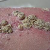
Unilateral Verrucous Psoriasis

Article Type
Changed
Sun, 02/07/2021 - 21:50


Case Report

An 80-year-old man with a history of hypertension and coronary artery disease presented to the dermatology clinic with a rash characterized by multiple asymptomatic plaques with overlying verrucous nodules on the left side of the abdomen, back, and leg (Figure 1). He reported that these “growths” appeared 20 years prior to presentation, shortly after coronary artery bypass surgery with a saphenous vein graft. The patient initially was given a diagnosis of verruca vulgaris and then biopsy-proven psoriasis later that year. At that time, he refused systemic treatment and was treated instead with triamcinolone acetonide ointment, with periodic surgical removal of bothersome lesions.

CT107002097_Fig1_AB.JPG
%3Cp%3E%3Cstrong%3EFigure%201.%20%3C%2Fstrong%3EVerrucous%20psoriasis%20on%20the%20left%20side%20of%20the%20body.%20A%2C%20Welldemarcated%2C%20scaly%2C%20erythematous%20plaques.%20B%2C%20Hyperkeratotic%20verrucous%20growths.%3C%2Fp%3E

At the current presentation, physical examination revealed many hyperkeratotic, yellow-gray, verrucous nodules overlying scaly, erythematous, sharply demarcated plaques, exclusively on the left side of the body, including the left side of the abdomen, back, and leg. The differential diagnosis included linear psoriasis and inflammatory linear verrucous epidermal nevus (ILVEN).

[embed:render:related:node:67427]

Skin biopsy showed irregular psoriasiform epidermal hyperplasia with acanthosis, hyperkeratosis, and papillomatosis, with convergence of the rete ridges, known as buttressing (Figure 2A). There were tortuous dilated blood vessels in the dermal papillae, epidermal neutrophils at the tip of the suprapapillary plates, and Munro microabscesses in the stratum corneum (Figure 2B). Koilocytes were absent, and periodic acid–Schiff staining was negative. Taken together, clinical and histologic features led to a diagnosis of unilateral verrucous psoriasis.

CT107002097_Fig2_AB.JPG
%3Cp%3E%3Cstrong%3EFigure%202.%3C%2Fstrong%3E%20Histopathology%20of%20verrucous%20psoriasis.%20A%2C%20Irregular%20psoriasiform%20epidermal%20hyperplasia%20with%20acanthosis%2C%20hyperkeratosis%2C%20papillomatosis%2C%20and%20buttressing%20(converging%20to%20the%20center)%20of%20rete%20ridges%20(H%26amp%3BE%2C%20original%20magnification%20%C3%9720).%20B%2C%20Tortuous%20dilated%20vessels%20were%20present%20on%20a%20biopsy%20specimen%20in%20dermal%20papillae%2C%20along%20with%20epidermal%20neutrophils%20that%20surmount%20the%20tips%20of%20suprapapillary%20plates.%20Intracorneal%20Munro%20microabscesses%20also%20were%20present%20(H%26amp%3BE%2C%20original%20magnification%20%C3%97100).%3C%2Fp%3E

Comment

Presentation and Histology
Verrucous psoriasis is a variant of psoriasis that presents with wartlike clinical features and overlapping histologic features of verruca and psoriasis. It typically arises in patients with established psoriasis but can occur de novo.

Histologic features of verrucous psoriasis include epidermal hyperplasia with acanthosis, papillomatosis, and epidermal buttressing.1 It has been hypothesized that notable hyperkeratosis observed in these lesions is induced by repeat trauma to the extremities in patients with established psoriasis or by anoxia from conditions that predispose to poor circulation, such as diabetes mellitus and pulmonary disease.1,2

Pathogenesis
Most reported cases of verrucous psoriasis arose atop pre-existing psoriasis lesions.3,4 The relevance of our patient’s verrucous psoriasis to his prior coronary artery bypass surgery with saphenous vein graft is unknown; however, the distribution of lesions, timing of psoriasis onset in relation to the surgical procedure, and recent data proposing a role for neuropeptide responses to nerve injury in the development of psoriasis, taken together, provide an argument for a role for surgical trauma in the development of our patient’s condition.

Treatment
Although verrucous psoriasis presents both diagnostic and therapeutic challenges, there are some reports of improvement with topical or intralesional corticosteroids in combination with keratolytics,3 coal tar,5 and oral methotrexate.6 In addition, there are rare reports of successful treatment with biologics. A case report showed successful resolution with adalimumab,4 and a case of erythrodermic verrucous psoriasis showed moderate improvement with ustekinumab after other failed treatments.7

Differential Diagnosis
Psoriasis typically presents in a symmetric distribution, with rare reported cases of unilateral distribution. Two cases of unilateral psoriasis arising after a surgical procedure have been reported, one after mastectomy and the other after neurosurgery.8,9 Other cases of unilateral psoriasis are reported to have arisen in adolescents and young adults idiopathically.

A case of linear psoriasis arising in the distribution of the sciatic nerve in a patient with radiculopathy implicated tumor necrosis factor α, neuropeptides, and nerve growth factor released in response to compression as possible etiologic agents.10 However, none of the reported cases of linear psoriasis, or reported cases of unilateral psoriasis, exhibited verrucous features clinically or histologically. In our patient, distribution of the lesions appeared less typically blaschkoid than in linear psoriasis, and the presence of exophytic wartlike growths throughout the lesions was not characteristic of linear psoriasis.

[embed:render:related:node:214538]

Late-adulthood onset in this patient in addition to the absence of typical histologic features of ILVEN, including alternating orthokeratosis and parakeratosis,11 make a diagnosis of ILVEN less likely; ILVEN can be distinguished from linear psoriasis based on later age of onset and responsiveness to antipsoriatic therapy of linear psoriasis.12

Conclusion

We describe a unique presentation of an already rare variant of psoriasis that can be difficult to diagnose clinically. The unilateral distribution of lesions in this patient can create further diagnostic confusion with other entities, such as ILVEN and linear psoriasis, though it can be distinguished from those diseases based on histologic features. Our aim is that this report improves recognition of this unusual presentation of verrucous psoriasis in clinical settings and decreases delays in diagnosis and treatment.

References
  1. Khalil FK, Keehn CA, Saeed S, et al. Verrucous psoriasis: a distinctive clinicopathologic variant of psoriasis. Am J Dermatopathol. 2005;27:204-207.
  2. Wakamatsu K, Naniwa K, Hagiya Y, et al. Psoriasis verrucosa. J Dermatol. 2010;37:1060-1062.
  3. Monroe HR, Hillman JD, Chiu MW. A case of verrucous psoriasis. Dermatol Online J. 2011;17:10.
  4. Maejima H, Katayama C, Watarai A, et al. A case of psoriasis verrucosa successfully treated with adalimumab. J Drugs Dermatol. 2012;11:E74-E75.
  5. Erkek E, Bozdog˘an O. Annular verrucous psoriasis with exaggerated papillomatosis. Am J Dermatopathol. 2001;23:133-135.
  6. Hall L, Marks V, Tyler W. Verrucous psoriasis: a clinical and histopathologic mimicker of verruca vulgaris. J Am Acad Dermatol. 2013;68(4 suppl 1):AB218.
  7. Curtis AR, Yosipovitch G. Erythrodermic verrucous psoriasis. J Dermatolog Treat. 2012;23:215-218.
  8. Kim M, Jung JY, Na SY, et al. Unilateral psoriasis in a woman with ipsilateral post-mastectomy lymphedema. Ann Dermatol. 2011;23(suppl 3):S303-S305.
  9. Reyter I, Woodley D. Widespread unilateral plaques in a 68-year-old woman after neurosurgery. Arch Dermatol. 2004;140:1531-1536.
  10. Galluzzo M, Talamonti M, Di Stefani A, et al. Linear psoriasis following the typical distribution of the sciatic nerve. J Dermatol Case Rep. 2015;9:6-11.
  11. Sengupta S, Das JK, Gangopadhyay A. Naevoid psoriasis and ILVEN: same coin, two faces? Indian J Dermatol. 2012;57:489-491.
  12. Morag C, Metzker A. Inflammatory linear verrucous epidermal nevus: report of seven new cases and review of the literature. Pediatr Dermatol. 1985;3:15-18.
Article PDF
Author and Disclosure Information

From the Department of Dermatology, Icahn School of Medicine at Mount Sinai, New York, New York.

The authors report no conflict of interest.

Correspondence: Riana D. Sanyal, MD, Icahn School of Medicine at Mount Sinai, 1 Gustave L. Levy Pl, New York, NY 10029 (riana.dutt@icahn.mssm.edu).

Issue
Cutis - 107(2)
Publications
Topics
Page Number
97-98
Sections
Author and Disclosure Information

From the Department of Dermatology, Icahn School of Medicine at Mount Sinai, New York, New York.

The authors report no conflict of interest.

Correspondence: Riana D. Sanyal, MD, Icahn School of Medicine at Mount Sinai, 1 Gustave L. Levy Pl, New York, NY 10029 (riana.dutt@icahn.mssm.edu).

Article PDF
Article PDF


Case Report

An 80-year-old man with a history of hypertension and coronary artery disease presented to the dermatology clinic with a rash characterized by multiple asymptomatic plaques with overlying verrucous nodules on the left side of the abdomen, back, and leg (Figure 1). He reported that these “growths” appeared 20 years prior to presentation, shortly after coronary artery bypass surgery with a saphenous vein graft. The patient initially was given a diagnosis of verruca vulgaris and then biopsy-proven psoriasis later that year. At that time, he refused systemic treatment and was treated instead with triamcinolone acetonide ointment, with periodic surgical removal of bothersome lesions.

CT107002097_Fig1_AB.JPG
%3Cp%3E%3Cstrong%3EFigure%201.%20%3C%2Fstrong%3EVerrucous%20psoriasis%20on%20the%20left%20side%20of%20the%20body.%20A%2C%20Welldemarcated%2C%20scaly%2C%20erythematous%20plaques.%20B%2C%20Hyperkeratotic%20verrucous%20growths.%3C%2Fp%3E

At the current presentation, physical examination revealed many hyperkeratotic, yellow-gray, verrucous nodules overlying scaly, erythematous, sharply demarcated plaques, exclusively on the left side of the body, including the left side of the abdomen, back, and leg. The differential diagnosis included linear psoriasis and inflammatory linear verrucous epidermal nevus (ILVEN).

[embed:render:related:node:67427]

Skin biopsy showed irregular psoriasiform epidermal hyperplasia with acanthosis, hyperkeratosis, and papillomatosis, with convergence of the rete ridges, known as buttressing (Figure 2A). There were tortuous dilated blood vessels in the dermal papillae, epidermal neutrophils at the tip of the suprapapillary plates, and Munro microabscesses in the stratum corneum (Figure 2B). Koilocytes were absent, and periodic acid–Schiff staining was negative. Taken together, clinical and histologic features led to a diagnosis of unilateral verrucous psoriasis.

CT107002097_Fig2_AB.JPG
%3Cp%3E%3Cstrong%3EFigure%202.%3C%2Fstrong%3E%20Histopathology%20of%20verrucous%20psoriasis.%20A%2C%20Irregular%20psoriasiform%20epidermal%20hyperplasia%20with%20acanthosis%2C%20hyperkeratosis%2C%20papillomatosis%2C%20and%20buttressing%20(converging%20to%20the%20center)%20of%20rete%20ridges%20(H%26amp%3BE%2C%20original%20magnification%20%C3%9720).%20B%2C%20Tortuous%20dilated%20vessels%20were%20present%20on%20a%20biopsy%20specimen%20in%20dermal%20papillae%2C%20along%20with%20epidermal%20neutrophils%20that%20surmount%20the%20tips%20of%20suprapapillary%20plates.%20Intracorneal%20Munro%20microabscesses%20also%20were%20present%20(H%26amp%3BE%2C%20original%20magnification%20%C3%97100).%3C%2Fp%3E

Comment

Presentation and Histology
Verrucous psoriasis is a variant of psoriasis that presents with wartlike clinical features and overlapping histologic features of verruca and psoriasis. It typically arises in patients with established psoriasis but can occur de novo.

Histologic features of verrucous psoriasis include epidermal hyperplasia with acanthosis, papillomatosis, and epidermal buttressing.1 It has been hypothesized that notable hyperkeratosis observed in these lesions is induced by repeat trauma to the extremities in patients with established psoriasis or by anoxia from conditions that predispose to poor circulation, such as diabetes mellitus and pulmonary disease.1,2

Pathogenesis
Most reported cases of verrucous psoriasis arose atop pre-existing psoriasis lesions.3,4 The relevance of our patient’s verrucous psoriasis to his prior coronary artery bypass surgery with saphenous vein graft is unknown; however, the distribution of lesions, timing of psoriasis onset in relation to the surgical procedure, and recent data proposing a role for neuropeptide responses to nerve injury in the development of psoriasis, taken together, provide an argument for a role for surgical trauma in the development of our patient’s condition.

Treatment
Although verrucous psoriasis presents both diagnostic and therapeutic challenges, there are some reports of improvement with topical or intralesional corticosteroids in combination with keratolytics,3 coal tar,5 and oral methotrexate.6 In addition, there are rare reports of successful treatment with biologics. A case report showed successful resolution with adalimumab,4 and a case of erythrodermic verrucous psoriasis showed moderate improvement with ustekinumab after other failed treatments.7

Differential Diagnosis
Psoriasis typically presents in a symmetric distribution, with rare reported cases of unilateral distribution. Two cases of unilateral psoriasis arising after a surgical procedure have been reported, one after mastectomy and the other after neurosurgery.8,9 Other cases of unilateral psoriasis are reported to have arisen in adolescents and young adults idiopathically.

A case of linear psoriasis arising in the distribution of the sciatic nerve in a patient with radiculopathy implicated tumor necrosis factor α, neuropeptides, and nerve growth factor released in response to compression as possible etiologic agents.10 However, none of the reported cases of linear psoriasis, or reported cases of unilateral psoriasis, exhibited verrucous features clinically or histologically. In our patient, distribution of the lesions appeared less typically blaschkoid than in linear psoriasis, and the presence of exophytic wartlike growths throughout the lesions was not characteristic of linear psoriasis.

[embed:render:related:node:214538]

Late-adulthood onset in this patient in addition to the absence of typical histologic features of ILVEN, including alternating orthokeratosis and parakeratosis,11 make a diagnosis of ILVEN less likely; ILVEN can be distinguished from linear psoriasis based on later age of onset and responsiveness to antipsoriatic therapy of linear psoriasis.12

Conclusion

We describe a unique presentation of an already rare variant of psoriasis that can be difficult to diagnose clinically. The unilateral distribution of lesions in this patient can create further diagnostic confusion with other entities, such as ILVEN and linear psoriasis, though it can be distinguished from those diseases based on histologic features. Our aim is that this report improves recognition of this unusual presentation of verrucous psoriasis in clinical settings and decreases delays in diagnosis and treatment.

References
  1. Khalil FK, Keehn CA, Saeed S, et al. Verrucous psoriasis: a distinctive clinicopathologic variant of psoriasis. Am J Dermatopathol. 2005;27:204-207.
  2. Wakamatsu K, Naniwa K, Hagiya Y, et al. Psoriasis verrucosa. J Dermatol. 2010;37:1060-1062.
  3. Monroe HR, Hillman JD, Chiu MW. A case of verrucous psoriasis. Dermatol Online J. 2011;17:10.
  4. Maejima H, Katayama C, Watarai A, et al. A case of psoriasis verrucosa successfully treated with adalimumab. J Drugs Dermatol. 2012;11:E74-E75.
  5. Erkek E, Bozdog˘an O. Annular verrucous psoriasis with exaggerated papillomatosis. Am J Dermatopathol. 2001;23:133-135.
  6. Hall L, Marks V, Tyler W. Verrucous psoriasis: a clinical and histopathologic mimicker of verruca vulgaris. J Am Acad Dermatol. 2013;68(4 suppl 1):AB218.
  7. Curtis AR, Yosipovitch G. Erythrodermic verrucous psoriasis. J Dermatolog Treat. 2012;23:215-218.
  8. Kim M, Jung JY, Na SY, et al. Unilateral psoriasis in a woman with ipsilateral post-mastectomy lymphedema. Ann Dermatol. 2011;23(suppl 3):S303-S305.
  9. Reyter I, Woodley D. Widespread unilateral plaques in a 68-year-old woman after neurosurgery. Arch Dermatol. 2004;140:1531-1536.
  10. Galluzzo M, Talamonti M, Di Stefani A, et al. Linear psoriasis following the typical distribution of the sciatic nerve. J Dermatol Case Rep. 2015;9:6-11.
  11. Sengupta S, Das JK, Gangopadhyay A. Naevoid psoriasis and ILVEN: same coin, two faces? Indian J Dermatol. 2012;57:489-491.
  12. Morag C, Metzker A. Inflammatory linear verrucous epidermal nevus: report of seven new cases and review of the literature. Pediatr Dermatol. 1985;3:15-18.
Issue
Cutis - 107(2)
Issue
Cutis - 107(2)
Page Number
97-98
Page Number
97-98
Publications
Publications
Topics
Article Type
Sections
Inside the Article

Practice Points

  • Verrucous psoriasis is a rare variant of psoriasis characterized by hypertrophic verrucous papules and plaques on an erythematous base.
  • Histologically, verrucous psoriasis presents with overlapping features of verruca and psoriasis.
  • Although psoriasis typically presents in a symmetric distribution, unilateral psoriasis can occur either de novo in younger patients or after surgical trauma in older patients.
Disallow All Ads
Content Gating
No Gating (article Unlocked/Free)
Alternative CME
Disqus Comments
Default
Use ProPublica
Hide sidebar & use full width
render the right sidebar.
Conference Recap Checkbox
Not Conference Recap
Clinical Edge
Display the Slideshow in this Article
Medscape Article
Article PDF Media
Image
Teambase ID
18001AFA.SIG
Disable zoom
Off