
BOSTON – Two award-winning researchers have received funding from the AGA Research Foundation to support their research into less invasive diagnostic techniques for GI patient care. The researchers shared updates on their findings at the 2016 AGA Tech Summit, which is sponsored by the AGA Center for GI Innovation and Technology.
Update on AGA-Boston Scientific Career Development Technology & Innovation Award
Guan Xu, Ph.D., of the University of Michigan, Ann Arbor, received the 2015 AGA–Boston Scientific award to support his investigation of an experimental, optically induced ultrasonic imaging technique called photoacoustic imaging, or PAI, in Crohn’s disease.
PAI uses pulsed laser light to penetrate tissues at different depths, allowing researchers to distinguish the absorption spectra indicative of the different chemical compositions seen in inflammatory and fibrous intestinal strictures.
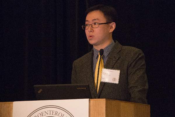
Guan Xu, Ph.D.
Though a number of experimental modalities are being investigated to characterize intestinal strictures less invasively, tissue biopsy is still considered the only reliable way to determine whether strictures are inflammatory or fibrous. Inflammatory strictures can be treated medically, while collagen-based fibrotic strictures must be removed surgically or treated with endoscopic dilation.
Dr. Xu joined Michigan’s radiology department as a postdoctoral research fellow in 2012 and is now a research investigator there. He previously helped develop a photoacoustic ultrasound imaging system for finger arthritis, designed for use in a clinical setting.
Dr. Xu and colleagues in the radiology, pathology, engineering and internal medicine departments at the University of Michigan are now investigating in animal models whether PAI is as accurate as tissue biopsy in distinguishing the two types of intestinal strictures seen in Crohn’s. They are also designing a capsule PAI device that can be fitted to an endoscope.
The PAI technology builds on and extends the ultrasound technology that is currently the standard of care, explained Dr. Xu, saying “We are standing on the shoulders of giants.”
At the Tech Summit, Dr. Xu presented an update of his research. Preliminary results have revealed significant differences in photoacoustic signal intensity among normal, inflammatory, and fibrotic bowel tissues in rats, and also strong correlations between photoacoustic images and histology in human stricture tissues. A prototype capsule PAI probe has been developed and it will soon be tested on mouse and rabbit models.
In an interview, Dr. Xu expressed “sincere gratitude to the AGA Research Foundation and Boston Scientific for the support of our preliminary study. We will work toward translation of our system to clinics.”
If Dr. Xu and his colleagues’ work is successful, clinicians may one day be able to distinguish between inflammatory and fibrotic intestinal strictures in Crohn’s disease patients without needing to perform endoscopic biopsy. The team’s findings may also prove useful in the preclinical investigation of antifibrotic medical therapies.
Update on AGA-Covidien Research & Development Pilot Award in Technology
Dr. David A. Katzka received the 2015 AGA–Covidien (now Medtronic) award, for his ongoing research into a simple, inexpensive technology as a possible alternative to repeat endoscopy and biopsy to monitor therapeutic response in people with eosinophilic esophagitis (EoE).
EoE is thought to result from exposure to food antigens, leading to inflammation and stricture formation. Current treatment recommendations include topical steroids and elimination diets, both of which are effective. However, response to withdrawal or reintroduction of problem foods often needs to be reassessed with repeat endoscopy and biopsy, which is both costly and time consuming.
Dr. Katzka, head of the Esophageal Interest Group at the Mayo Clinic in Rochester, Minn., has been developing the Cytosponge, an easily swallowed encapsulated mesh that expands in the stomach. Upon retrieval (by means of an attached string), the sponge provides a sample of esophageal mucosa. The technology was originally developed as a screening modality for esophageal cancer and Barrett’s esophagus. Unlike with endoscopy, no sedation is required.
A preliminary feasibility study in 20 patients with EoE who underwent both Cytosponge and endoscopic biopsy found good correlation of eosinophils between the two technologies, and a high sensitivity (84%) in diagnosing EoE. Genetic analysis of patient specimens obtained with Cytosponge showed consistency with previous transcriptome data seen on biopsy specimens. Importantly, patients favored the Cytosponge as faster and easier to tolerate than endoscopy.
Dr. Katzka’s research team is now studying a larger patient group to compare the Cytosponge with endoscopic biopsy as a reliable and independent means of monitoring disease activity and response to therapy. Genomic analysis is also being performed on specimens so that as molecular predictors of response to therapy are further developed and become available, these tests may be also be performed – with the idea of limiting or eliminating the need for endoscopy.
