Clinical Challenges

Clinical Challenges - November 2016
What’s your diagnosis?

What’s your diagnosis?

By Benjamin Kloesel, MD, Vishal S. Chandan, MD, and Glenn L. Alexander, MD. Published previously in Gastroenterology (2012;143:1439, 1692).

A 30-year-old woman with a past medical history of hypothyroidism presents for evaluation of epigastric discomfort, nausea without emesis, abdominal bloating, and watery, nonbloody diarrhea for 5 months. This was associated with a 15-pound weight loss. Complete blood count, liver function tests, thyroid-stimulating hormone, immunoglobulin (Ig) levels, and IgG/IgA tissue transglutaminase (tTG) were within normal limits. Stool studies for bacterial pathogens, Giardia, Clostridium difficile toxin, and ova/parasites were negative.

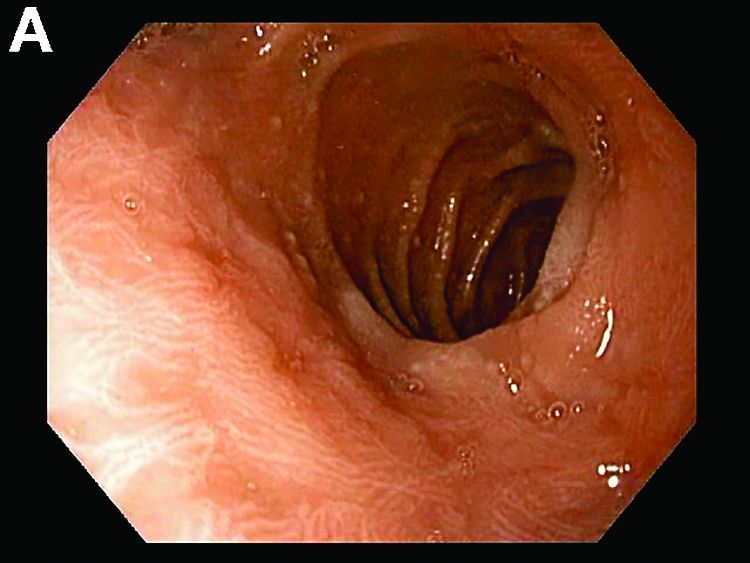
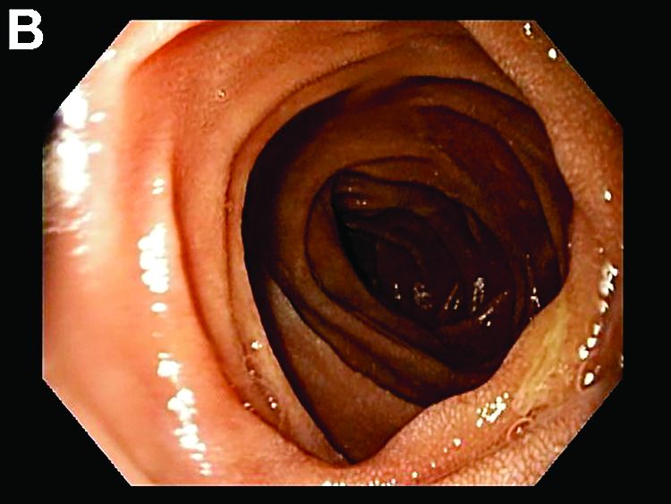
An upper endoscopy revealed minimal antral erythema and abnormal duodenal bulb (Figure A) with a normal-appearing postbulbar duodenum (Figure B). Biopsies of the stomach, duodenal bulb, and second portion of the duodenum were obtained.
What is the diagnosis?


Answer to “What’s your diagnosis?” on page X: Collagenous gastritis and collagenous sprue
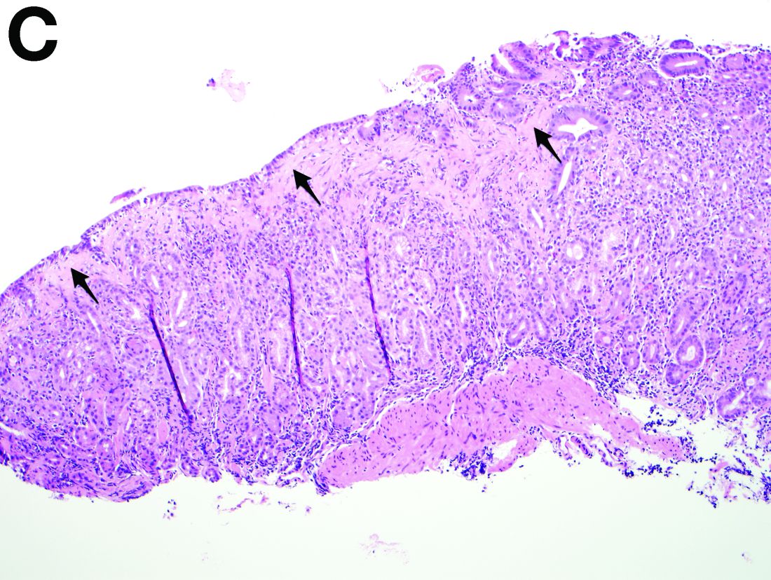
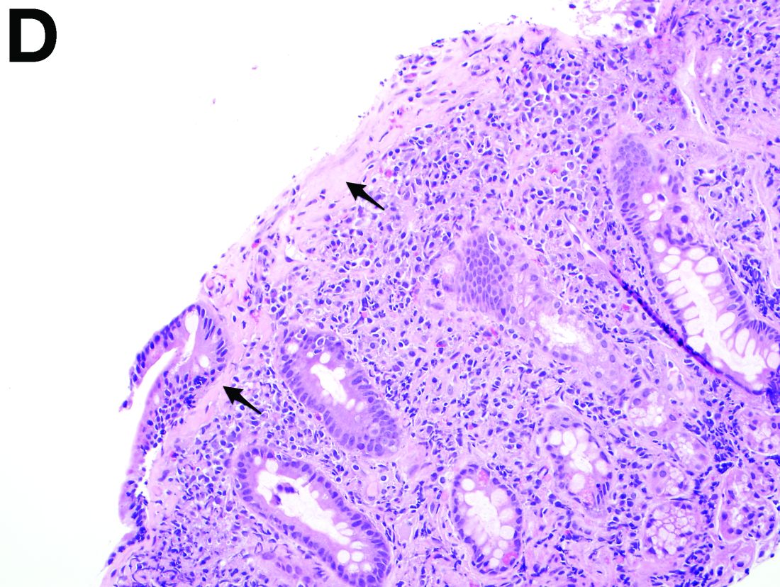
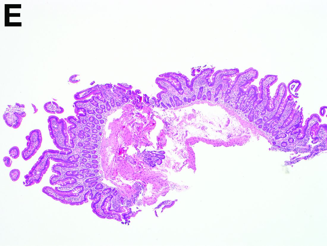
Histology revealed a thickened subepithelial collagen band in the stomach (Figure C) and total villous blunting with an increased subepithelial collagen band in the duodenal bulb (Figure D). Conversely, the histologic appearance of the second portion of the duodenum was completely normal (Figure E). Gastric biopsies were negative for Helicobacter pylori. The diagnoses of collagenous gastritis and collagenous sprue were established. Treatment included initiation of a gluten-free diet along with a trial of omeprazole.
Collagenous gastritis is an uncommon condition first described in 1989. A recent review defined two patient groups based on age and presentation with children and young adults presenting with anemia and abdominal pain and older adults predominantly presenting with diarrhea. In the adult group, two cases were associated with celiac disease, whereas an additional five individuals were found to have concurrent collagenous or lymphocytic colitis.1 Various treatment approaches including acid suppression, corticosteroids, misoprostol, mesalamine, and sucralfate were tried with mostly disappointing results; interventions therefore mainly focus on concurrent diseases such as gluten-free diet for celiac disease and budesonide for collagenous colitis.2
Despite a sensitivity in the 90%-98% range of tTG antibodies for the detection of celiac disease, this case demonstrates that a negative IgA/IgG tTG cannot entirely exclude the diagnosis and that duodenal biopsies should still be obtained in the setting of a high clinical suspicion for celiac disease or an abnormal-appearing duodenum on endoscopy.

Recent studies have also shown the importance of obtaining at least 1 biopsy from the duodenal bulb to avoid missing the diagnosis of celiac disease. In 126 patients with newly established celiac disease and 85 patients with a previous diagnosis on a gluten-free diet presenting for reevaluation, villous atrophy was limited to the duodenal bulb in 9% and 14% of cases, respectively.3

References

1. Brain, O., Rajaguru, C., Warren, B. et al. Collagenous gastritis: reports and systematic review. Eur J Gastroenterol Hepatol. 2009;21:1419-24.

2. Gopal, P., McKenna, B.J. The collagenous gastroenteritides: similarities and differences. Arch Pathol Lab Med. 2010;134:1485-9.

3. Evans, K.E., Aziz, I., Cross, S.S. et al. A prospective study of duodenal bulb biopsy in newly diagnosed and established adult celiac disease. Am J Gastroenterol. 2011;106:1837-742.

Next Article: