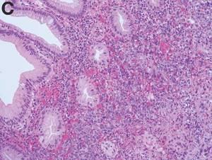
Clinical Challenges

Clinical Challenges - July 2016
What's Your Diagnosis?

What's Your Diagnosis?

By Dr. Maw-Soan Soon, Dr. Chih-Jung Chen, and Dr. Hsu-Heng Yen. Published previously in Gastroenterology (2012:143:1155, 1400).

A 35-year-old woman was referred for evaluation of intermittent abdominal pain for 3 months.

She denied weight loss or change in bowel habits.

A stool fecal occult test was positive and colonoscopy was performed (Figure A).

After colonoscopy, further studies with abdominal computed tomography (CT; Figure B) revealed additional findings.


The diagnosis

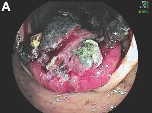
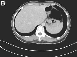
The colonoscopy disclosed an erythematous, ulcerated tumor with spontaneous bleeding in the ascending colon (Figure A). Noncontrast abdominal CT showed multiple hypoattenuating lesions in the liver. These lesions had central enhancement in the early arterial phase and progressive enhancement in the delayed phase (Figure B).

The colon biopsy revealed proliferation of atypical cells admixed with blood-filled spaces in the lamina propria (Figure C; stain: H&E; original magnification, 200 ×). The tumor cells were strongly and diffusely positive for CD31 (Figure D; stain: H&E; original magnification, 200 ×). The liver biopsy showed the same findings. A diagnosis of primary colonic angiosarcoma with liver metastasis was made. The patient was referred to an oncologist for further treatment.

Angiosarcoma is a rare, malignant tumor arising from vascular endothelium. These tumors usually develop in skin and in the soft tissue of the head and neck region.1 Gastrointestinal angiosarcomas are rare with fewer than 20 cases reported in the literature. 1,2 Patients with colonic angiosarcoma usually present with abdominal pain, rectal bleeding, or a palpable abdominal mass. 1,2 On colonoscopy, the tumor has been described as a submucosal tumor 1,2or a hemorrhagic mass with spontaneous bleeding 3, as in this case.

Colonic angiosarcomas usually progress rapidly with a poor prognosis. Patients with older age (more than 60 years), large tumor size (more than 5 cm), or metastatic disease have a poorer prognosis.1,2 Fewer than 10% of the patients survive 2 years after the diagnosis. The treatment is usually operative resection. The role of adjuvant chemotherapy is unclear.References

1. Lo, T.H., Tsai, M.S., Chen, T.A. Angiosarcoma of sigmoid colon with intraperitoneal bleeding: case report and literature review. Ann R Coll Surg Engl. 2011;93:e91-3.

2. El Chaar, M., McQuay, N. Jr. Sigmoid colon angiosarcoma with intraperitoneal bleeding and early metastasis. J Surg Educ. 2007;64: 54-6.

3. Cammarota, G., Schinzari, G., Larocca, L.M. et al. Duodenal metastasis from a primary angiosarcoma of the colon. Gastrointest Endosc. 2006;63:330.

Next Article: