Practical Teaching Cases

A Rare Endoscopic Clue to a Common Clinical Condition

Published previously in Gastroenterology (2017;152:492-3)


AGA Institute
A 64-year-old woman presented to a local emergency department after noting large-volume passage of bright red blood from her colostomy site over several days. She denied any associated abdominal pain, recent changes in bowel pattern, nausea, vomiting, orthostatic symptoms, abdominal trauma, NSAID use, or recent manipulation of the ostomy concurrent with her symptoms. Her past medical history was significant for hypertension and remote stage 1B cervical cancer complicated by radiation-induced enteritis, proctitis, and terminal ileal stricture. Four years prior to her current presentation, surgical resection of the terminal ileum had been performed with a side-to-side ileoascending colostomy and creation of an end-sigmoid colostomy for management of persistent diarrhea and fecal incontinence.


AGA Institute
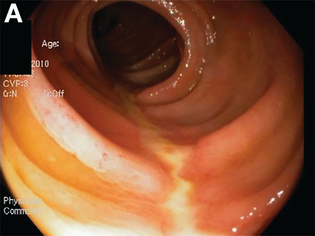
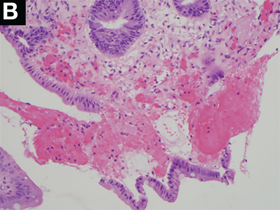
On examination, the patient was mildly hypotensive (BP 100/65 mm Hg) with bright red blood visible in the ostomy bag. Laboratory testing revealed normal hemoglobin (15 g/dL) and an upright abdominal x-ray showed changes consistent with her prior surgical history. Because of ongoing ostomy bleeding, the patient was transferred to a tertiary facility where repeat labs now showed mild anemia (hemoglobin 13 g/dL). A colonoscopy demonstrated unilateral linear ulceration of the distal transverse colon, measuring 5 cm long and 8 mm in diameter with a clean white base (Figure A). The remaining colonic mucosa was unremarkable except for scattered diverticula within the transverse colon. Biopsies obtained from the ulcer showed foci of cryptitis, focal fibrosis, and hemorrhage within the lamina propria (Figure B).

Dr. Anderson and Dr. Sweetser are in the Division of Gastroenterology and Hepatology, Mayo Clinic College of Medicine, Rochester, Minn.

Which of the following is the most likely cause for the patient’s symptoms?

Diverticular disease–associated colitis

Cytomegalovirus colitis

Colonic ischemia

Radiation colitis

The correct answer is C: colonic ischemia.

AGA Institute

Figure A

The endoscopic findings are notable for colon single-stripe sign (CSSS), which is a highly specific feature of colonic ischemia (Figure A). The diagnosis of colon ischemia is further supported by the histologic features of cryptitis, focal fibrosis, and hemorrhage within the lamina propria (Figure B). In this case, the patient’s history of radiation exposure and hypotension were both likely predisposing factors for colonic hypoperfusion and subsequent colon ischemia. With conservative medical therapy, the patient experienced complete resolution of symptoms.

AGA Institute

Figure B

Diverticular disease-associated colitis (answer A) is less likely given the lack of interdiverticular mucosal involvement and linear ulceration pattern, which also contrasts with the deep, “punched-out” appearance typically associated with ulceration of cytomegalovirus colitis (answer B). The endoscopic findings associated with chronic radiation colitis (answer D) characteristically include evidence of mucosal scarring, friability, and scattered angioectasias. The CSSS was initially described as a manifestation of colonic ischemia by Zuckerman et al. who hypothesized the linear nature of this lesion likely reflected segmental vascular compromise.1 Concordant with the presented case, of the 26 patients with CSSS included in the Zuckerman et al. study, all had a stripe measuring 5 cm in length while a minority (4 patients) had transverse colon involvement.1 Also in parallel with this case, others have reported successful nonoperative management of patients with CSSS in the setting of ischemia.1,2 Overall, the comparatively favorable outcome in patients with CSSS compared to those with circumferential colonic ischemia suggests this finding may reflect a more mild form of disease.1 n

References

1. Zuckerman G.R., et al. Am J Gastroenterol. 2003;98:2018-22.

2. Tanapanpanit O., Pongpirul K. BMJ Case Rep. 2015 Sept. 17;2015.

This article has an accompanying continuing medical education activity, also eligible for MOC credit (see gastrojournal.org for details). Learning Objective: Upon completion of this activity, successful learners will be able to recognize colon single-stripe sign as an endoscopic feature of colonic ischemia.

Next Article: