Authors’ Disclosure Statement: The authors report no actual or potential conflict of interest in relation to this article.
Dr. Frank is Assistant Professor, Department of Orthopaedic Surgery, University of Colorado School of Medicine, Aurora, Colorado. Mr. Golijanin is a Medical Student, Geisel School of Medicine, Dartmouth Medical School, Hanover, New Hampshire. Dr. Vopat is Assistant Professor, Department of Orthopaedic Surgery, University of Kansas, Kansas City, Kansas. Dr. Gross is a Resident, DGMD Medical, Omaha, Nebraska. Dr. Chauhan is a Resident, Ninewells Hospital, Dundee, United Kingdom. Dr. Romeo is Chief of Orthopaedics, Rothman Institute New York, New York. Dr. Provencher is an Orthopaedic Surgeon, The Steadman Clinic, Vail, Colorado.
Address correspondence to: Rachel M. Frank MD, Department of Orthopaedic Surgery, University of Colorado School of Medicine, Aurora, CO 80045 (email, Rachel.Frank@ucdenver.edu).
Am J Orthop. 2018;47(6). Copyright Frontline Medical Communications Inc. 2018. All rights reserved.
Rachel M. Frank, MD Petar Golijanin, BS Bryan G. Vopat, MD Daniel J. Gross, MD Vidhya Chauhan, BS Anthony A. Romeo, MD CAPT Matthew T. Provencher, MD, MC, USNR . Impact of Sagittal Rotation on Axial Glenoid Width Measurement in the Setting of Glenoid Bone Loss. Am J Orthop.
June 5, 2018
References
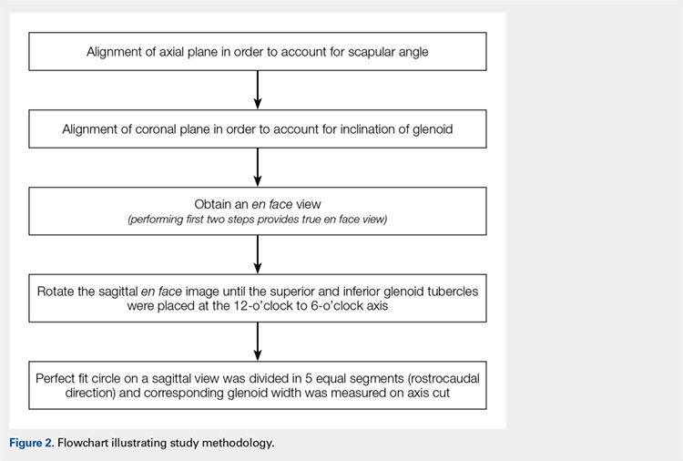
IMAGE ANALYSIS AND REFORMATTING
In a de-identified fashion, all CT scans were imported and analyzed using open-source Digital Imaging and Communications in Medicine (DICOM) software (OsiriX MD, version 2.5.1 64-bit). By following a previously developed method, CT scans were reformatted using OsiriX MPR. The OsiriX software has an MPR function that allows simultaneous manipulation of 2-D CT scans in 3 orthogonal planes: axial, sagittal, and coronal. In the MPR mode, the alternation of 1 plane directly affects the orientation of the remaining 2 planes. Thus, by using an MPR, one can analyze the impact that a default CT scan performed relative to the gantry of the table, UNCORR, has on the axial images.
First, the en face view was obtained via a 2-step process: alignment of the axial plane to account for the scapular angle, followed by alignment of the coronal plane to adjust for the glenoid inclination.15 These 2 adjustments provided a true en face sagittal glenoid view. The final adjustment step was a sagittal en face rotation of the glenoid such that the superior and inferior glenoid tubercles were placed on the 12-o’clock to 6-o’clock axis (CORR scan). Previous studies have identified a central longitudinal axis that was used in this method to align the supraglenoid tubercle with the 12-o’clock to 6-o’clock axis on the glenoid face.15,17,18 The standard error of mean was 1.21°. This new CORR view resulted in axial cuts through the glenoid that were oriented perpendicular to the 12-o’clock to 6-o’clock axis. The UNCORR and CORR images were assessed in the axial plane at 5 standardized cuts and measured for AP glenoid width by 2 independent observers in a blinded, randomized fashion. When the measured AP width of the UNCORR scan was less than that measured on the CORR scan, the AP width of the glenoid was considered underestimated, and the degree of GBL was considered overestimated (Figure 2).
SCAPULAR ANGLE
Scapular angle measurements were performed on the axial view as the angle between a line through the long axis of the body of the scapula, and a line parallel to the CT gantry table.15,19 Subsequently, the axial plane was aligned to the glenoid surface.
CORONAL INCLINATION
Coronal inclination measurements were performed on the sagittal view as the angle between a line tangential to the face of the glenoid and a line perpendicular to the CT gantry table. Positive values represented superior inclination, while negative values represented inferior glenoid inclination.15
SAGITTAL ROTATION
Sagittal rotation measurements were performed using the built-in angle measurement tool in OsiriX in the sagittal plane since the degree of rotation required aligning the long axis of the glenoid to the 12-o’clock to 6-o’clock axis. The amount of rotation was defined as the rotation angle.15