

A) Globular enlarged uterus INCORRECT
A homogeneously enlarged uterus in the absence of fibroids is characteristic of adenomyosis.1
Transvaginal pelvic ultrasound demonstrates a homogenously enlarged uterus (arrows) without evidence of a focal fibroid.
B) Cystic myometrial spaces INCORRECT
Myometrial cysts are dilated cystic glands or foci of hemorrhage within heterotopic endometrial tissue.2 They are often less than 5 mm in size but can be extensive and of variable sizes and can be differentiated from arcuate veins seen in the outer myometrium by the use of color Doppler.1,2
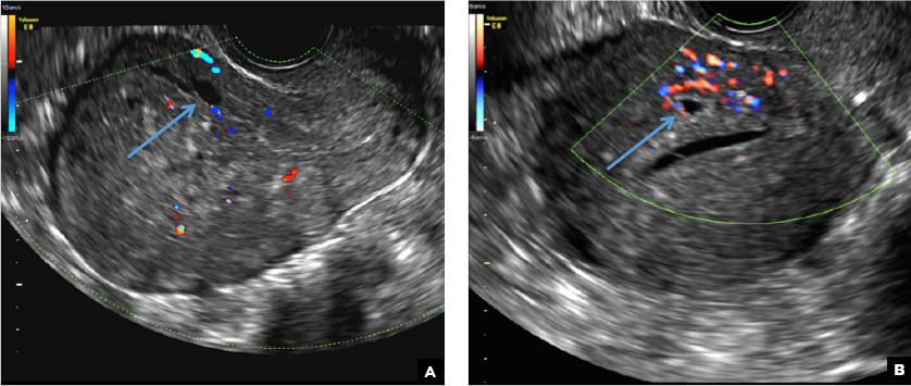
Transvaginal pelvic ultrasound on 2 different patients demonstrates well-defined anechoic cysts in the myometrium without vascular flow on color Doppler (long arrows).
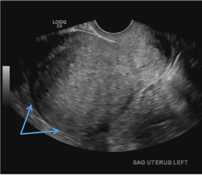
C) Asymmetric myometrial thickening INCORRECT
The finding of asymmetric uterine wall thickening in adenomyosis is usually seen when there is focal disease and presents with wall thickening that demonstrates anteroposterior asymmetry.1
(A) Transvaginal pelvic ultrasound demonstrates asymmetric wall thickening of the posterior myometrium (long arrow). (B) Transvaginal pelvic ultrasound of a different patient demonstrates asymmetric wall thickening of the anterior myometrium (long arrow).
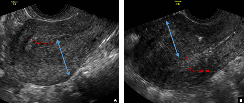
D) Indistinct endomyometrial interface INCORRECT
The heterotopic endometrial tissue invading the myometrium obscures the normal endometrial myometrial border with pseudo-widening of the endometrial echo resulting in poor definition of the endomyometrial junction.1,2
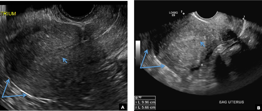
Transvaginal pelvic ultrasounds from 2 different patients demonstrate poor definition of the endomyometrial interface (short arrows) in addition to the globular appearance of the uterus (long arrows).
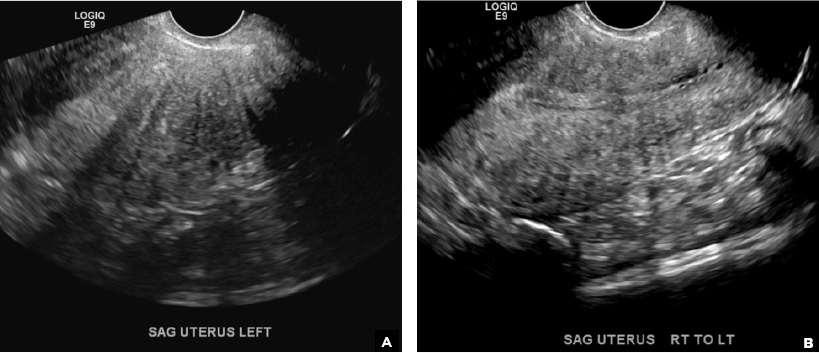
E) Myometrial linear striations CORRECT
A hyperplastic reaction to the infiltration of the heterotrophic endometrial glands into the myometrium results in radiating linear echogenic striations (sometimes referred to as the "venetian blinds" sign).1,2
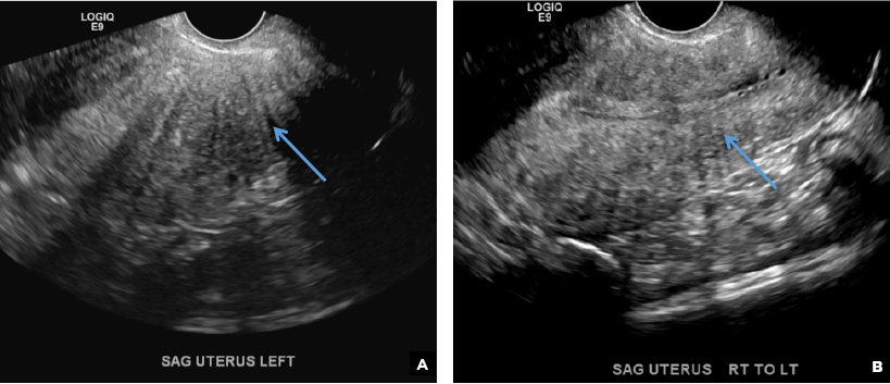
Transvaginal pelvic ultrasound images demonstrate myometrial linear echogenic striations (long arrows) radiating from the anterior uterine wall.