From the Editor

Benefits and pitfalls of open power morcellation of uterine fibroids
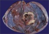
The current practice of open power morcellation is being scrutinized by those within and outside of the ObGyn community. We need to re-examine our...

Letters from Readers
How I avoid open power morcellation
Several gynecologic surgeons at my hospital (I am one of them) have developed approaches to hysterectomy and myomectomy that avoid open power morcellation.
For laparoscopic hysterectomy, we place two or three 5-mm ports and use a retroperitoneal approach. We identify the ureters in the retroperitoneal space and trace them into the pelvis. They often need to be lateralized in cases involving endometriosis or large fibroids. We then coagulate the uterine artery where it crosses over the ureter, as the vessel is smaller and straighter in this location. This strategy reduces blood loss and lessens the need to fulgurate the vessels at the cervix, where the ureter is vulnerable to injury.
When the uterus is large, we morcellate it transvaginally or through a 3-cm transverse suprapubic incision after placing a retractor. Be aware that 60% of women with fibroids can have coexisting adenomyosis. As Dr. Barbieri noted in his editorial, use of an internal morcellator can disperse small tissue fragments throughout the peritoneal cavity, where they may “re-implant” and undergo malignant transformation.
For laparoscopic myomectomy, I place 600 µg of misoprostol—a very potent drug used to contract the uterus, with a large safety margin—in the rectum before starting the surgery, to help reduce blood loss. I also place atraumatic vascular clips on the uterine vessels in the retroperitoneum and on the utero-ovarian ligaments. Along with the use of vasopressin, these techniques markedly reduce blood loss. A 3-cm transverse suprapubic incision then is used, with fibroids removed through a retractor and the uterus then repaired.
Traditional laparoscopy enables the surgeon to change the degree of Trendelenburg, which is very helpful.
Our patients almost always go home within several hours. On postoperative day 2, most of them report needing only nonsteroidal anti-inflammatory drugs for pain and say they are eating and ambulating well. In my experience, these patients appear to have much less postoperative pain than those who undergo vaginal hysterectomy.
In 2012, 97% of major gynecologic surgical cases at my hospital were minimally invasive, with a very low complication rate. The procedures included resection of severe endometriosis, removal of very large uteri, myomectomy, and treatment of endometrial cancer, including node dissection.
Ray Wertheim, MD
Director of the AAGL Center of Excellence Minimally Invasive Gynecology Program,
Inova Fair Oaks Hospital, Fairfax, Virginia
Morcellated leiomyosarcoma is a very real risk
As a gynecologic oncologist at the University of Miami, I saw at least three cases (referred to us) of morcellated leiomyosarcomas. Within 1 or 2 months of the original surgery, each patient had to be returned to the operating room (OR) for an exploration, and disseminated tumor was found. No patient survived beyond 1 year. Therefore, I was surprised at the 5-year survival rate of 46% presented in Dr. Barbieri’s editorial.
Karen Nishida, MD
Miramar, Florida
Dr. Barbieri responds
I appreciate Dr. Wertheim’s excellent surgical pearls. Clinicians at the Inova hospitals are national leaders in advancing women’s health.
I agree with Dr. Nishida that open power morcellation of an occult leiomyosarcoma likely worsens the prognosis of the patient. Even with intensive treatment some of these patients will die within 2 years from sarcomatosis.
Related Articles:
Benefits and pitfalls of open power morcellation of uterine fibroids Robert L. Barbieri, MD (Editorial, February 2014)
Options for reducing the use of open power morcellation of uterine tumors Robert L. Barbieri, MD (Editorial, March 2014)
A likely case of encephalitis linked to teratoma
I enjoyed the interesting case presented by Dr. Barbieri and Dr. Clark in their editorial on ovarian teratoma and encephalitis. A few months ago a neurologist called me about a similar case. The patient was experiencing seizures and had a 17-cm mass beneath the umbilicus. I agreed to resect the mass, which was suspicious for an immature teratoma, and the patient recovered slowly.
I am glad to see this reference in the literature now. I am sure the editorial by Dr. Barbieri and Dr. Clark will prove handy to our colleagues.
Courtney Ridley, MD
Sacramento, California
Dr. Barbieri responds
I appreciate Dr. Ridley taking time from her busy practice to write about her experience with an ovarian tumor and encephalitis. The practice of obstetrics and gynecology is very rewarding because we are always encountering new ideas and constantly evolving our approaches to patient care. The link between encephalitis and ovarian teratoma was discovered only in 2007. It is regrettable that many women likely died before 2007 because they did not undergo timely removal of an ovarian teratoma, a simple operation.

The current practice of open power morcellation is being scrutinized by those within and outside of the ObGyn community. We need to re-examine our...
The most promising alternative to open power morcellation is morcellation in a bag, described here

Can resection of an ovarian teratoma reverse this patient’s disabling brain disease?

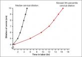
We are in a new era. Our patients, and their labors, have changed on a global scale. To optimally manage labor you need to use these new norms in...
