Breast density is a strong, prevalent, and potentially modifiable risk factor for breast cancer, which makes it of special interest to clinicians whose jobs involve breast cancer risk prediction. That was the theme of a talk by Karla Kerlikowske, MD, of the UCSF Helen Diller Family Comprehensive Cancer Center in San Francisco. Dr. Kerlikowske delivered the John I. Brewer Memorial Lecture May 3 at the 2015 Annual Clinical Meeting of the American College of Obstetricians and Gynecologists in San Francisco.
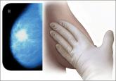
Mammographic breast density is a radiologic term, Dr. Kerlikowske explained. “The only way to really know someone’s breast density is if they have a mammogram.” The whiter the mammogram, the denser the breast. The darker the mammogram, the fattier the breast.
According to the American College of Radiology, the following 4 categories of breast composition are defined by the “visually estimated” content of fibroglandular-density tissue within the breasts:
A. The breasts are almost entirely fatty.
B. There are scattered areas of fibroglandular density.
C. The breasts are heterogeneously dense, which may obscure small masses.
D. The breasts are extremely dense, which lowers the specificity of mammography.
Categories C and D signify dense breasts, which contain a high degree of collagen, epithelial cells, and stroma. In the United States, more than 25 million women are thought to have dense breasts.
Women who have a family history of breast cancer are more likely to have dense breasts. And women who have dense breasts have an elevated risk of breast cancer. They also have a higher risk of advanced disease, as well as a higher risk of large, high-grade, and lymph node-positive tumors, said Dr. Kerlikowske.
Twenty-two states now have laws mandating that women found to have heterogeneously dense or extremely dense breasts be notified of their status, said Dr. Kerlikowske. That prompts the question: How should these patients be managed?
Breast density declines with age. Breast density also is influenced by body mass index (BMI). As BMI increases, density declines.
Breast density also can be affected by medications, such as hormone therapy and tamoxifen, Dr. Kerlikowske said.
For example, breast density declines about 1% to 2% per year in postmenopause. In postmenopausal women who take estrogen alone, breast density increases slightly. “But the real increase is for people who take estrogen plus progestin,” said Dr. Kerlikowske. “It’s thought that the progestin component is what increases breast density.” Estrogen-progestin therapy confers the same risk of breast cancer as that faced by a premenopausal woman with dense breasts.
As for tamoxifen, it reduces breast density by 2% to 3% per year in postmenopausal women, Dr. Kerlikowske said. “People who have a decrease of more than 10% in breast density are those who have a reduction in breast cancer.” If a woman doesn’t have that reduction with tamoxifen—about half of women don’t—there is no reduction in breast cancer mortality.
“There’s some thought that you should look at mammograms during the first year of tamoxifen use and, if you don’t see a change, consider switching to another medication,” said Dr. Kerlikowske.
More frequent mammograms and supplemental imaging are options for detecting cancers early. Among the modalities that have been studied in this regard are ultrasonography, tomosynthesis, and breast magnetic resonance imaging (MRI).
“If you do more tests, such as ultrasound, you will definitely find additional lesions,” said Dr. Kerlikowske. “There’s no question. But what are the harms?”
The biopsy rate almost doubles after ultrasonography, compared with mammography. And the number needed to screen to detect cancer is fairly high. For mammography, that number is about 250. For ultrasonography, tomosynthesis, and breast MRI, it is higher.
Tomosynthesis is more cost-effective than supplemental ultrasonography because it decreases the number of false positives, Dr. Kerlikowske said.
Not every woman with dense breasts is at high risk for breast cancer, said Dr. Kerlikowske. And although breast density is prevalent, it is potentially modifiable.
Nevertheless, breast density confers an elevated risk of breast cancer and can also mask tumors. Women with dense breasts likely should avoid the use of postmenopausal hormone therapy. They also may be candidates for more frequent mammography and/or supplemental imaging.
The Breast Cancer Surveillance Consortium (BCSC) Risk Calculator is the only tool that incorporates breast density. In the works is a new model that also incorporates benign breast disease.
A risk-prediction tool from the Breast Cancer Surveillance Consortium (BCSC) is the only model to incorporate breast density. The BCSC Risk Calculator is available free of charge for the iPhone and iPad (an Android version is in the works). The tool takes 5 factors into consideration in estimating a woman’s 5-year risk of developing invasive breast cancer:
• age
• race/ethnicity
• breast density
• family history of breast cancer (first-degree relative)
• personal history of breast biopsy.
The tool is designed for use by health professionals. It is not appropriate for determining risk in women younger than 35 years or older than 79 years; women with a previous diagnosis of breast cancer, lobular carcinoma in situ, ductal carcinoma in situ, or atypical ductal hyperplasia; or women who have undergone breast augmentation. Other risk-prediction models are more appropriate for women with a BRCA mutation.