
Established criteria for identifying inflammatory back pain in people with ankylosing spondylitis do not perform well in identifying axial involvement in people with psoriatic arthritis and neither does clinical judgment, a study shows.
 feellife/Thinkstock
There’s reason to believe that the natural history of patients with psoriatic arthritis (PsA) who have axial disease could differ from those without it, and there are differences in how well criteria that are currently used to identify inflammatory back pain (IBP) in people with ankylosing spondylitis (AS) perform in people with PsA, study first author Kristy S. Yap, MBBS, and her colleagues at the University of Toronto Psoriatic Arthritis Clinic wrote in Annals of the Rheumatic Diseases.
“Axial involvement in PsA is a marker of disease severity, and those with axial disease often have worse outcomes, compared with peripheral arthritis alone,” they wrote.
This is backed up by European League Against Rheumatism recommendations that advise clinicians to consider prescribing tumor necrosis factor inhibitors for people with PsA who have active axial involvement.
“Thus, an important question when evaluating a patient with PsA is to determine if axial PsA is present,” they wrote, noting that it was currently unclear whether the three sets of criteria that exist for defining inflammatory back pain in AS – Calin, Rudwaleit, and Assessment of Spondyloarthritis International Society (ASAS) – were useful for screening for axial involvement in people with PsA.
The researchers therefore set out to determine the agreement between rheumatologist judgment of the presence of IBP as well as the presence of IBP according to the three criteria in 171 patients with PsA (52% male, average age 46.6 years), 96 of whom reported chronic back pain, including 65 with IBP and 31 with nonspecific back pain.
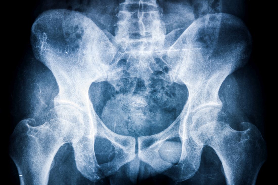
Radiology data from these patients showed that 27 with baseline x-rays fulfilled the New York radiographic criteria for AS, and 45 had radiographic sacroiliitis not satisfying NY criteria (excluding grade 1) and/or syndesmophytes. Nine out of 31 patients with no axial disease on x-ray had evidence of axial disease on MRI. Eighteen out of 54 patients had axial involvement without back pain.
Results showed that agreement (kappa coefficient) between rheumatologist judgment of IBP and IBP criteria in patients with back pain was moderate and was highest for the Calin criteria (0.70; 95% confidence interval, 0.56-0.85), followed by the ASAS criteria (0.61; 95% CI, 0.46-0.76) and the Rudwaleit criteria (0.59; 95% CI, 0.44-0.74).
When x-ray or MRI change was considered “gold standard” for axial involvement for all patients, the specificity was high for rheumatologist judgment of IBP as well as Calin, Rudwaleit, and ASAS criteria, but their sensitivity was low, the researchers reported.
When the investigators compared positive likelihood ratios (LRs) for the presence of back pain, the Rudwaleit criteria (2.17) performed the best in ruling in axial disease, whereas the LRs were 1.75 for Calin and 1.86 for ASAS criteria. Rheumatologist-reported back pain (0.68) performed the best for ruling out axial disease when comparing negative LRs.
“The low positive LRs of the Calin, Rudwaleit, and ASAS criteria as well as that of rheumatologist report of back pain or judgment of IBP for [axial] PsA defined as any axial radiological change found in our study suggests that none of these criteria performed well in detecting axial disease in patients with PsA,” the study authors wrote.