From the Department of Dermatology, Icahn School of Medicine at Mount Sinai, New York, New York.
Drs. Lanoue and Chen report no conflict of interest. Dr. Goldenberg is a speaker for, has received honoraria from, and has performed research and consulting for LEO Pharma; PharmaDerm; and Valeant Pharmaceuticals International, Inc.
Correspondence: Gary Goldenberg, MD, Department of Dermatology, 5 E 98th St, 5th Floor, New York, NY 10029 (garygoldenbergmd@gmail.com).
Field cancerization is the process in which a singular cell accumulates genetic mutations following carcinogen exposure and then divides to create a “field” of monoclonal premalignant cells. In this study, microscopically identified actinic keratoses (AKs) were used as markers of field cancerization in all excision specimens of squamous cell carcinomas (SCCs), basal cell carcinomas (BCCs), and malignant melanomas (MMs) received by our institution’s dermatopathology department over a 3- to 6-month period. Our findings provide additional evidence for the theory of field cancerization, its association with cutaneous malignancies, and the need to assess the extent of field damage when determining treatment strategies.
Clinically apparent and subclinical actinic keratoses usually are present in patients, a concept known as field cancerization, and it is important to treat both types of lesions.
Actinic keratoses are present in the field of cutaneous malignancies, including basal cell carcinoma, squamous cell carcinoma, and melanoma.
References
The concept of field cancerization was first proposed in 1953 by Slaughter et al1 in their study of oral squamous carcinomas. Their findings of multifocal patches of premalignant disease, abnormal tissue surrounding tumors, multiple localized primary tumors, and tumor recurrence following surgical resection was suggestive of a field of dysplastic cells with malignant potential.1 Since then, modern molecular techniques have been used to establish a genetic basis for this model in many different types of cancer, including cutaneous malignancies.2-4 The field begins from a singular stem cell, which undergoes one or more genetic changes that allow for a growth advantage compared to surrounding cells. The stem cell then divides, forming a patch of clonal daughter cells that displace the surrounding normal epithelium. Growth of this patch eventually leads to a dysplastic field of monoclonal cells, which notably does not yet show invasive growth or metastatic behavior. Over time, continued carcinogenic exposure results in additional genetic alterations among different cells in the field, which leads to new subclonal proliferations that share common clonal origin but exhibit unique genetic changes. Eventually, transformative events may occur, resulting in cells with invasive and metastatic properties, thus forming a carcinoma.5
In the case of cutaneous malignancies, UV radiation in the form of UVA and UVB rays is the most common source of carcinogenesis. It is well established that UV radiation has numerous effects on the body, including but not limited to local and systemic immunosuppression, alteration of signal transduction pathways, and the development of mutations in DNA via direct damage by UVB or indirect damage by free radical formation with UVA.6,7 Normally, DNA is protected from UV radiation–induced genetic alteration by the p53 gene, TP53. As such, damage to this gene is highly associated with cancer induction. One study found that more than 90% of squamous cell carcinomas (SCCs) and more than 50% of basal cell carcinomas (BCCs) contain UV-like mutations in TP53.8 The concept of field cancerization suggests that because the skin surrounding cutaneous malignancies has been exposed to the same chronic UV light as the initial lesion, it is at an increased risk for genetic abnormalities and thus possible malignant transformation.
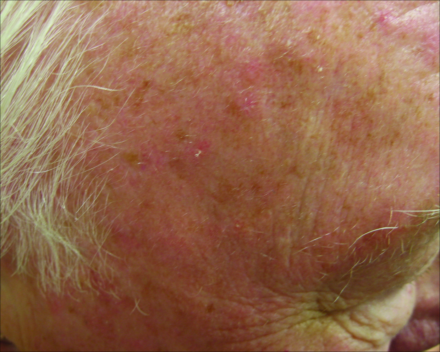
Actinic keratoses (AKs) are common neoplasms of the skin that generally are regarded as precancerous lesions or may be considered to be the earliest stage of SCC in situ.9 Actinic keratoses usually develop as a consequence of chronic exposure to UV radiation and often are clinically apparent as erythematous scaly papules in sun-exposed areas (Figure 1).10 They also are identified histologically as atypical keratinocytes along the basal layer of the epidermis with possible enlargement, hyperchromatic nuclei, lack of maturation, mitotic figures, inflammatory infiltrate, and/or hyperkeratosis.10 Furthermore, the genetic changes associated with AKs are well documented and are strongly associated with changes to p53.11 Given these characteristics, AKs serve as good markers of genetic damage with potential for malignancy. In this study, we used histologically identified AKs to assess the presence of field damage in the tissue immediately surrounding excision specimens of SCCs, BCCs, and malignant melanomas (MMs).
Figure 1. Scaly erythematous papules typical of actinic keratosis.
Methods
This study was approved by the Program for the Protection of Human Subjects at the Icahn School of Medicine at Mount Sinai (New York, New York) prior to initiation. All cutaneous specimens submitted to the dermatopathology service for consultation between April 2013 and June 2013 were reviewed for inclusion in this study. Data collection was extended for MMs to include all specimens from January 2013 to June 2013 given the limited number of cases in the original data collection period.
Initial screening for this study was done electronically and assessed for a diagnosis of SCC (Figure 2), BCC (Figure 3), or MM (Figure 4) as determined by a board-certified dermatopathologist (G.G.). The resulting pool of specimens was then screened to include only excision specimens and to exclude curettage specimens and superficial specimens that lacked dermis. In this study, we chose to look at reexcisions rather than initial biopsies so that there was a greater likelihood of having an intact epidermis surrounding a malignancy that could be assessed for the presence of AKs as markers for field cancerization. Specimens were examined in full via serial transverse cross-sections at 3-mm intervals. Additional step sections were obtained at smaller intervals when margins were close or unclear.
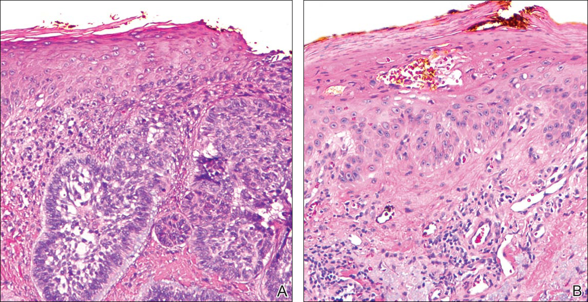
Figure 2. On the left, an actinic keratosis demonstrates atypical keratinocytes along the basal layer with hyperchromatic nuclei and atypical maturation. Separated by a section of histologically normal epithelium, a squamous cell carcinoma is seen on the right (H&E, original magnification ×200).
Figure 3. Residual basal cell carcinoma (A)(H&E, original magnification ×200). Actinic keratosis from the same excision with notable parakeratosis and solar elastosis in the dermis (B)(H&E, original magnification ×200).
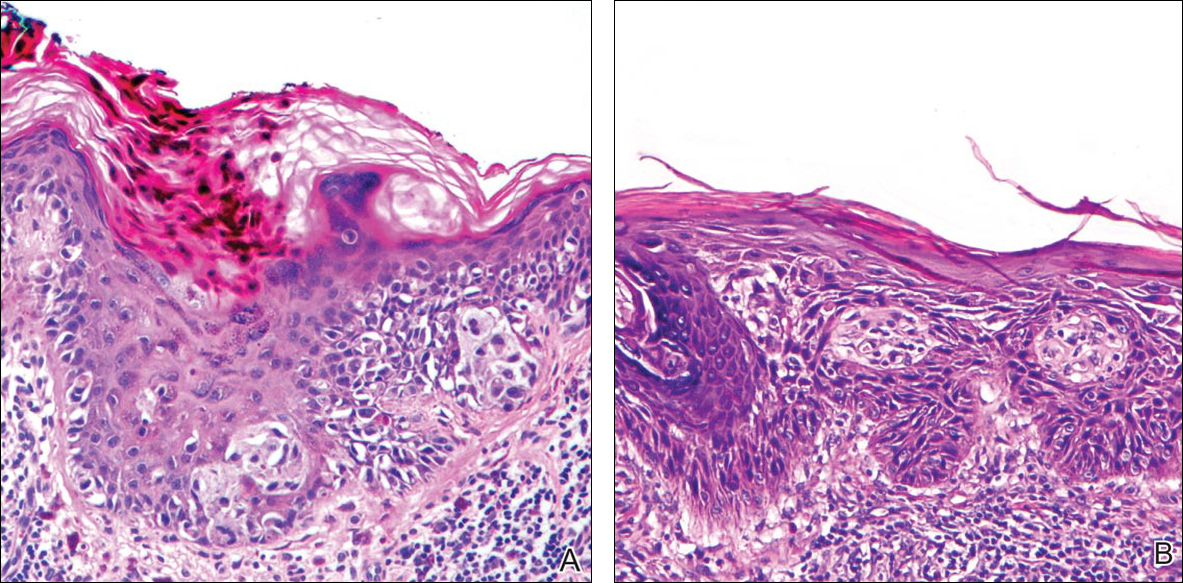
Figure 4. Malignant melanoma (A)(H&E, original magnification ×200). Incidental actinic keratosis in the same excision specimen, both images exhibit a lymphocytic infiltrate.