Hand-foot-and-mouth disease (HFMD) is a viral illness caused by several enteroviruses, most commonly coxsackievirus A16 (CVA16) and enterovirus 71 (EV71). The disease is generally seen in children younger than 5 years, characterized by lesions of the oral mucosa, palms, and soles, usually lasting 7 to 10 days. Other coxsackie type A viruses, including CVA6, CVA9, and CVA10, also are associated with HFMD.1-5 Although CVA16 has traditionally been the primary strain causing HFMD, CVA6 has become a major cause of HFMD outbreaks in the United States and worldwide in recent years.6-12 Interestingly, CVA6 also has been found to be associated with adult HFMD, which has increased in incidence. The CVA6 strain was first identified in association with the disease during HFMD outbreaks in Finland and Singapore in 2008,13,14 with similar strains detected in subsequent outbreaks in Taiwan, Japan, Spain, France, China, India, and the United States.12,15-25 Most cases took place in warmer months, with one winter outbreak in Massachusetts in 2012.24
Herein, we review the incidence of CVA6, as well as its atypical presentation, diagnosis, and treatment to aid dermatologists. Given the increasing incidence of HFMD caused by CVA6 and its often atypical presentation, it is important for dermatologists to be aware of this increasingly notable disease state and its viral cause.
Incidence of CVA6
Coxsackievirus A6 has been identified as the cause of many reported outbreaks of HFMD since it was first identified in 2008, and it is known to cause both pediatric and adult outbreaks.7-12 It may even be surpassing other strains in frequency in certain areas. In Tianjin, China, for example, EV71 and CVA16 were the most common serotypes causing HFMD from 2008 to 2012; however, in 2013, CVA6 was the most prevalent strain.26 According to one study, “[n]early every Chinese city showed a sharp rise in [CVA6].”27
The incidence of CVA6 also has been increasing in other areas.28 In Spain, CVA6 overtook CVA16 as the dominant cause of HFMD during 2011 and 2012 outbreaks.29 From 2011 to 2012, there was a CVA6-associated HFMD outbreak in North America, with 63 cases reported to the Centers for Disease Control and Prevention (CDC), including 15 adult cases, with approximately 50% having been exposed to children with HFMD.9 In 2014, a Minnesota college with approximately 1000 students reported 9 suspected cases of HFMD to the Minnesota Department of Health. Coxsackievirus A6 was isolated, sequenced, and identified by the CDC in 5 of 9 patients (age range, 19–47 years).9
In 2015, an outbreak of HFMD took place at Lackland Air Force Base in Texas during a basic military training. Eight cases were confirmed and 45 cases were suspected. The rate of infection was 0.4% (50/12,270) among trainees and 0.3% (2/602) among instructors.7 Eight of 12 nasopharyngeal swabs tested positive for EV by way of local real-time reverse transcription–polymerase chain reaction (RT-PCR). Four nasopharyngeal swabs were sent to the CDC for evaluation and all were positive for CVA6.7
Presentation
Because the prevalence of CVA6 has increased, it is important to be able to identify the presentation of HFMD caused by this strain. Coxsackievirus A6 has been found to affect a broader demographic and cause more severe cases of HFMD with its unique constellation of findings compared to other known strains. Patients present with flulike symptoms; higher fever than present in typical HFMD; and a longer duration of disease, typically lasting 2 weeks. Patients also may present with more severe skin disease compared to classic HFMD, not only including vesicles but also large bullae, erosions, and ulcers on the dorsal and plantar feet (Figure 1). Skin lesions often are painful and spread to a wider distribution than typical of HFMD, which can include the face, proximal extremities, lips, perianal and groin skin, scalp, and dorsal feet and hands (Figure 2). These areas are classically spared in the prototypical presentation of HFMD in children.2,6,24,30-33 Vesicles that are typically football shaped (Figure 3) are a diagnostic clue of the disease. After patients have recovered from the disease, they can have delayed-onset palmar and plantar desquamation that usually presents 1 to 3 weeks after the disease. Additionally, another postsyndrome finding is onychomadesis, or detachment of the nail plate from the nail matrix.6,34-37 This process likely occurs due to direct cytopathic effect to the nail matrix from the viral infection.24,37 Blistering may be severe and can form hemorrhagic bullae.24 Although cutaneous findings are more severe, neurologic involvement actually is more rare in the CVA6 strain compared to other viral strains known to cause HFMD, specifically EV71. One study found only 2.4% of 141 patients infected with CVA6 had central nervous system involvement, specifically aseptic meningitis or encephalitis.21,24
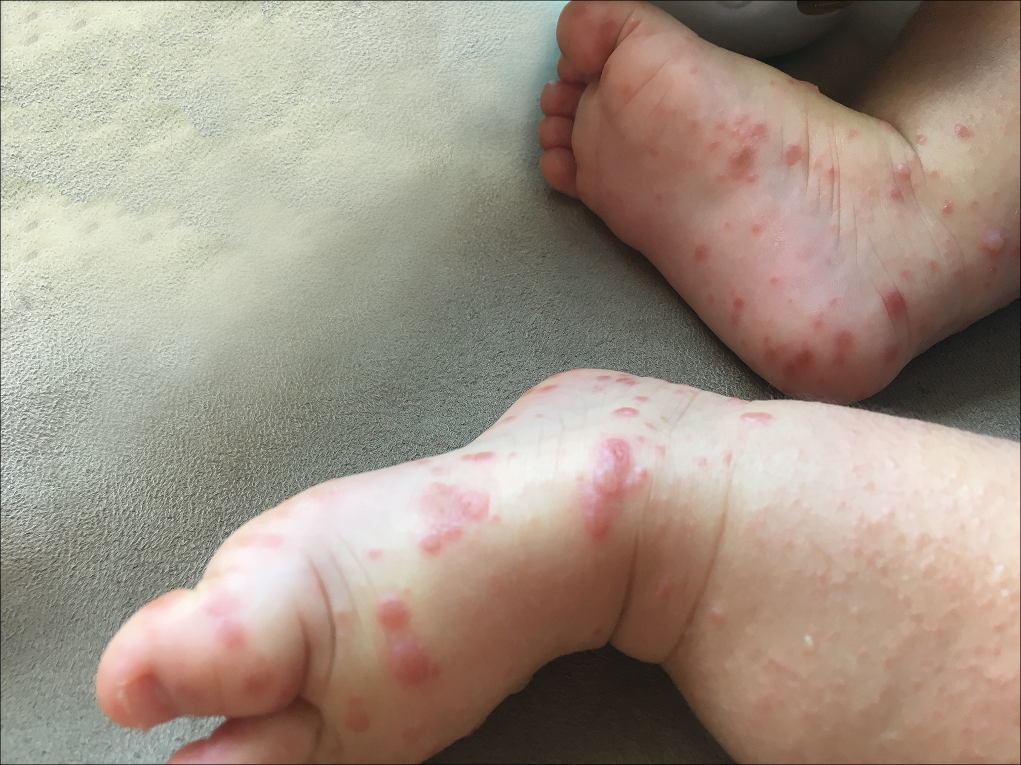 Photograph courtesy of Lauren Snitzer, MD (Houston, Texas).
Figure 1. Numerous vesicles on an erythematous base and erythematous papules on the dorsal and plantar feet.
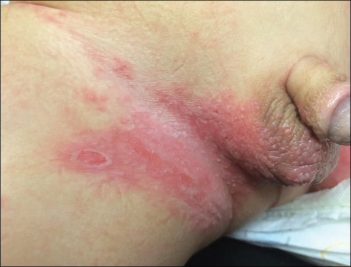 Photograph courtesy of Lauren Snitzer, MD (Houston, Texas).
Figure 2. Ill-defined, erythematous, eroded plaque on the right proximal thigh, inguinal fold, and right scrotum.
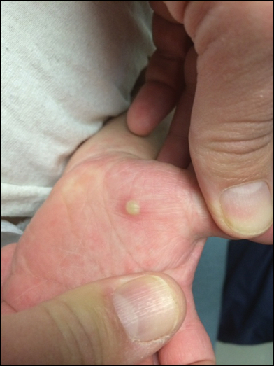 Photograph courtesy of Lauren Snitzer, MD (Houston, Texas).
Figure 3. Classic football-shaped lesion of hand-foot-and-mouth disease.
In patients with atopic dermatitis, CVA6 also shows a predilection to appear in areas of skin disease, such as the flexural regions of the arms and legs, and is referred to as eczema coxsackium.24,38,39 It can mimic eczema herpeticum or varicella superinfection, which are important considerations to include in the differential diagnosis. Additionally, CVA6-induced lesions often show up in previously irritated or traumatized areas such as sunburns, fungal infections, and diaper dermatitis in children. Lesions have been described to sometimes mimic Gianotti-Crosti syndrome, with involvement of the extensor surfaces, buttocks, and cheeks, and sparing of the trunk.24