Dr. Cahn is from the Memorial Sloan Kettering Cancer Center, New York, New York. Dr. Elston is from the Department of Dermatology and Dermatologic Surgery, Medical University of South Carolina, Charleston.
The authors report no conflict of interest.
The eTable is available in the Appendix online at www.mdedge.com/dermatology.
Correspondence: Brian A. Cahn, MD, 1275 York Ave, New York, NY 10065 (briancahn1489@gmail.com).
Sponges from the phylum Porifera exist throughout the world in marine and freshwater environments. Although many encounters with humans are benign, some may lead to local dermatologic manifestations and in rare cases can cause more severe systemic reactions. Initial decontamination is of utmost importance to diminish the severity of the reaction. As contact between humans and coastal environments increases, it is important for physicians to know how to recognize and treat sponge dermatitis.
Sponges exist in both marine and freshwater environments throughout the world.
Immediate management of sponge dermatitis should include decontamination by removing the sponge spicules with tape or rubber cement followed by dilute vinegar soaks.
Topical steroids may be used only after initial decontamination. Use of oral steroids may be needed for more severe reactions.
References
Sponges are among the oldest animals on earth, appearing more than 640 million years ago before the Cambrian explosion, a period when most major animal phyla appeared in the fossil records.1 More than 10,000 species of sponges have been identified worldwide and are distributed from polar to tropical regions in both marine (Figure 1) and freshwater (Figure 2) environments. They inhabit both shallow waters as well as depths of more than 2800 m, with shallower sponges tending to be more vibrantly colored than their deeper counterparts. The wide-ranging habitats of sponges have led to size variations from as small as 0.05 mm to more than 3 m in height.2 Their taxonomic phylum, Porifera (meaning pore bearers), is derived from the millions of pores lining the surface of the sponge that are used to filter planktonic organisms.3 Flagellated epithelioid cells called choanocytes line the internal chambers of sponges, creating a water current that promotes filter feeding as well as nutrient absorption across their microvilli.4 The body walls of many sponges consist of a collagenous skeleton made up of spongin and spicules of silicon dioxide (silica) or calcium carbonate embedded in the spongin connective tissue matrix.5 Bath sponges lack silica spicules.
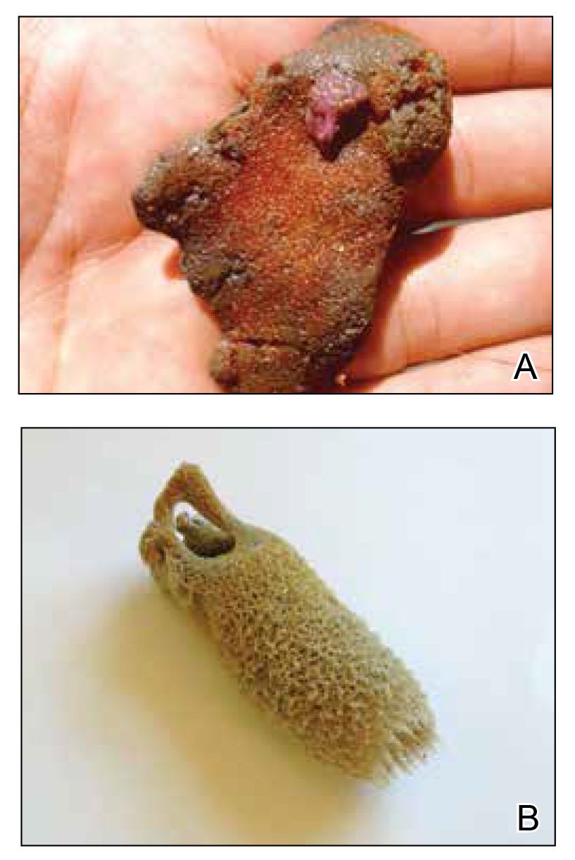
Figure 1. Marine sponges. A, Tedania ignis (fire sponge). Photograph courtesy of Vidal Haddad Jr, MD, PhD (Botucatu, São Paulo, Brazil). B, Agelas conifera (brown tube sponge). Photograph courtesy of Dirk M. Elston, MD (Charleston, South Carolina).
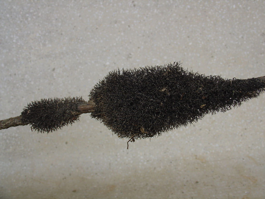
Figure 2. Cauxi sponge, a type of freshwater sponge. Photograph courtesy of Vidal Haddad Jr, MD, PhD (Botucatu, São Paulo, Brazil).
Sponges have been used in medicine for centuries. The first use in Western culture was recorded in 405 bce in The Frogs, a comedy by Aristophanes in which a sponge was placed on a character’s heart following a syncopal episode. Additionally, in many Hippocratic writings, the use of sponges is outlined in the treatment of a variety of ailments. Similarly, the ancient Chinese and Greeks used burnt sponge and seaweed as a source of iodine to treat goiters.6,7 Modern research focuses on the use of sponge metabolites for their antineoplastic, antimicrobial, and anti-inflammatory effects.8 Identification of spongouridine and spongothymidine from the sponge Tectitethya crypta led to the development of cytarabine and gemcitabine8 as well as the discovery of the antiviral agent vidarabine.9 The monoclonal antibody assay for the detection of shellfish poisoning was prepared using the sponge Halichondria okadai.10
Mechanisms and Symptoms of Injury
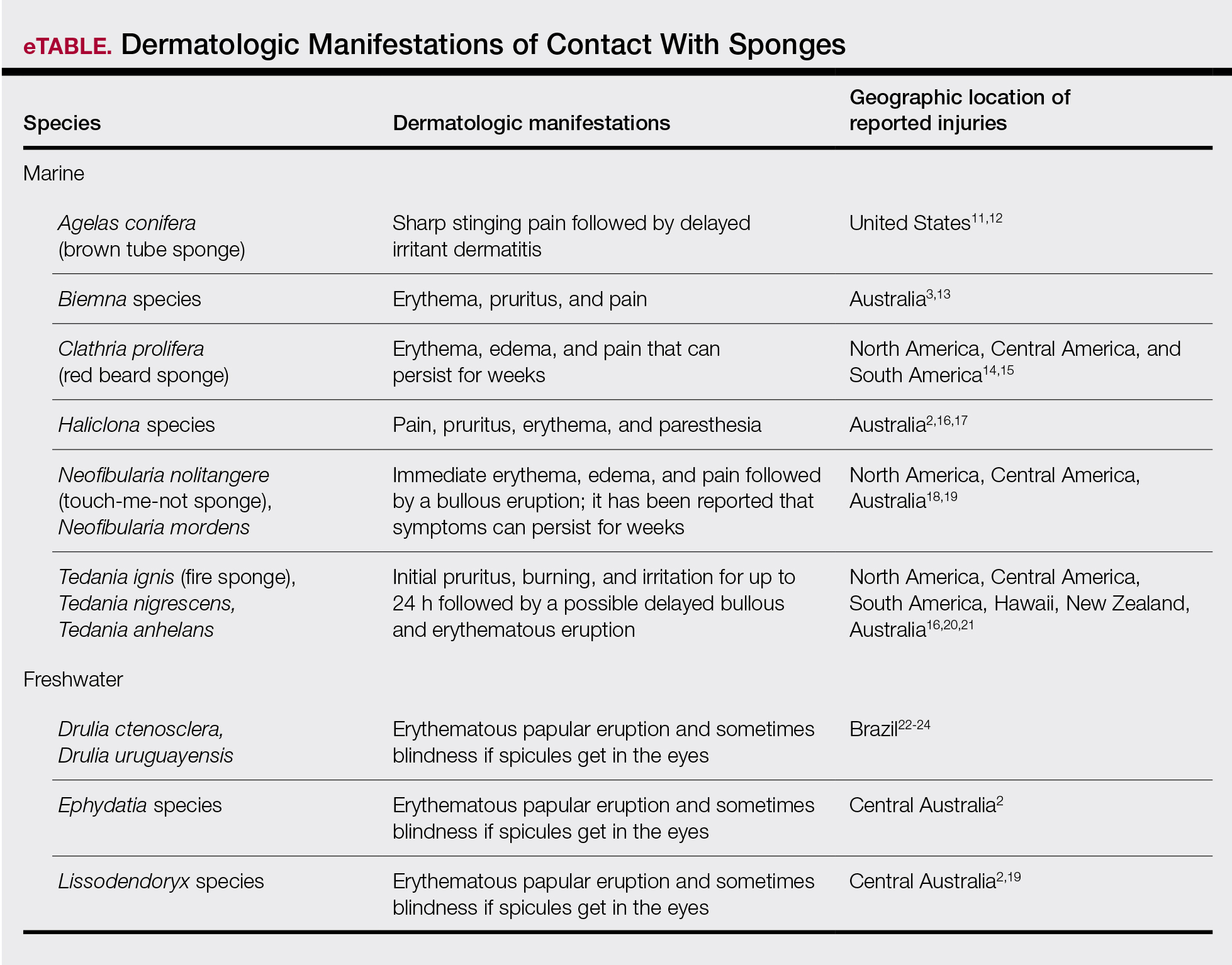
Bathing sponges (silk sponges) derived from Spongia officinalis are harmless. Other sponges can exert their damaging effects through a variety of mechanisms that lead to dermatologic manifestations (eTable). Some species of sponges produce and secrete toxic metabolites (eg, crinotoxins) onto the body surface or into the surrounding water. They also are capable of synthesizing a mucous slime that can be irritating to human skin. Direct trauma also can be caused by fragments of the silica or calcium carbonate sponge skeleton penetrating the skin. Stinging members of the phylum Cnidaria can colonize the sponge, leading to injury when a human handles the sponge.25-27
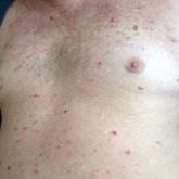
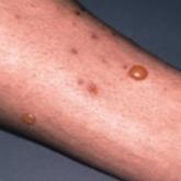
Sponge dermatitis can be divided into 2 major categories: an initial pruritic dermatitis (Figure 3) that occurs within 20 minutes to a few hours after contact and a delayed irritant dermatitis caused by penetration of the spicules and chemical agents into skin.28 Importantly, different species can lead to varying manifestations.
Figure 3. Initial pruritic eczematous plaques with erythema and edema after handling a toxic marine sponge. Photograph courtesy of Vidal Haddad Jr, MD, PhD (Botucatu, São Paulo, Brazil).
The initial pruritic dermatitis is characterized by itching and burning that progresses to local edema, vesiculation, joint swelling, and stiffness. Because most contact with sponges occurs with handling, joint immobility may ensue within 24 hours of the encounter. Rarely, larger areas of the skin are affected, and fever, chills, malaise, dizziness, nausea, purulent bullae, muscle cramps, and formication may occur.28 Anaphylactic reactions have been described in a small subset of patients. There have even been reports of delayed (ie, 1–2 weeks following exposure) erythema multiforme, livedo reticularis, purpura, and dyshidrotic eczema.16,20,29 The irritant dermatitis caused by spicule trauma is due to a foreign body reaction that can be exacerbated by toxins entering the skin. In severe cases, desquamation, recurrent eczema, and arthralgia can occur.30 In general, more mild cases should self-resolve within 3 to 7 days. Dermatologic conditions also can be caused by organisms that inhabit sponges and as a result produce a dermatitis when the sponge is handled, including sponge divers disease (maladie des plongeurs), a necrotic dermatitis caused by stinging Cnidaria species.31 Dogger Bank itch, first described as a dermatitis caused by sensitization to (2-hydroxyethyl) dimethylsulfoxonium chloride, initially was isolated from the sea chervil (a type of Bryozoan); however, that same chemical also was later found in sponges, producing the same dermatitis after handling the sponge.32 Freshwater sponges also have been reported to be injurious and exist worldwide. In contrast to marine sponges, lesions from freshwater sponges are disseminated pruritic erythematous papules with ulcerations, crusts, and secondary infections.22 The disseminated nature of the dermatitis caused by freshwater sponges is due to contact with the spicules of dead sponges that are dispersed throughout the water rather than from direct handling. Sponge dermatitis occurs mostly in sponge collectors, divers, trawlers, and biology students and has been reported extensively in the United States, Caribbean Islands, Australia, New Zealand, and Brazil.18,27,33,34
Management
Treatment should consist of an initial decontamination; the skin should be dried, and adhesive tape or rubber cement should be utilized to remove any spicules embedded in the skin. Diluted vinegar soaks should be initiated for 10 to 30 minutes on the affected area(s) 3 or 4 times daily.19 The initial decontamination should occur immediately, as delay may lead to persistent purulent bullae that may take months to heal. Topical steroids may be used following the initial decontamination to help relieve inflammation. Antihistamines and nonsteroidal anti-inflammatory drugs may be used to alleviate pruritus and pain, respectively. Severe cases may require systemic glucocorticoids. Additionally, immunization status against tetanus toxoid should be assessed.35 In the event of an anaphylactic reaction, it is important to maintain a patent airway and normalized blood pressure through the use of intramuscular epinephrine.36 Frequent follow-up is warranted, as serious secondary infections can develop.37 Patients also should be counseled on the potential for delayed dermatologic reactions, including erythema multiforme. Contact between humans and coastal environments has been increasing in the last few decades; therefore, an increase in contact with sponges is to be expected.22