Article

A Rare Case of Segmental Neurofibromatosis With Multiple Blue-Red Pseudoatrophic Plaques
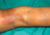
We report the case of a 5-year-old girl who presented with 2 blue-red atrophic plaques on the left leg as well as subcutaneous nodules that were...
Sandra Beverly, MD; Elizabeth Foley, MD; Dori Goldberg, MD
From the Department of Medicine, University of Massachusetts Medical School, Worcester.
The authors report no conflict of interest.
Correspondence: Sandra Beverly, MD, C/O Dori Goldberg, MD, Department of Medicine, University of Massachusetts Medical School, Hahnemann Campus, 281 Lincoln St, Worcester, MA 01605 (sandy.beverly@gmail.com).

Cutaneous neurofibromas are a clinical feature of both neurofibromatosis type I and neurofibromatosis type II. Neurofibromas are discrete masses that arise from peripheral nerves and are composed of Schwann cells, mast cells, fibroblasts, and perineural cells. Current treatment of neurofibromas consists primarily of standard surgical excision or laser therapy. A removal technique that is time efficient, cost effective, and poses little discomfort or functional impairment for the patient is desirable. The authors describe the technique of shave removal with electrodesiccation for multiple neurofibromas.
To the Editor:
Cutaneous neurofibromas are a clinical feature of both neurofibromatosis type I (NF-1) and neurofibromatosis type II (NF-2). Neurofibromatosis type I occurs in 1 in 3000 live births and NF-2 occurs in 1 in 40,000 live births.1 Neurofibromas are discrete masses that arise from peripheral nerves and are composed of Schwann cells, mast cells, fibroblasts, and perineural cells. They commonly appear during or after puberty in the majority of patients and increase in number as patients age.1,2 Cutaneous neurofibromas can be painful and pruritic as well as cosmetically unappealing. Current treatment of neurofibromas consists primarily of standard surgical excision or laser therapy. Reports of loop electrocoagulation in the operating room, combination erbium:YAG and CO2 laser, and Nd:YAG laser treatments of neurofibromas have been published in the literature.3-5 These treatments can be effective, but the bothersome lesions in patients with neurofibromatosis often are multiple, making these procedures time consuming and costly. A removal technique that is time efficient, cost effective, and poses little discomfort or functional impairment for the patient is desirable. We describe the technique of shave removal with electrodesiccation for multiple neurofibromas.
A 24-year-old woman with NF-1 presented to the dermatology clinic for treatment of several pruritic neurofibromas of the trunk, including the bilateral breasts (Figure, A). We performed shave removal of each lesion with electrodesiccation of the lesion base with successful treatment of symptoms and acceptable cosmetic outcome (Figure, B). The patient identified symptomatic and cosmetically bothersome neurofibromas. The lesions were disinfected with an alcohol swab and anesthetized with 1% lidocaine and a 1:200,000 dilution of epinephrine. A shave biopsy blade was used to remove each neurofibroma at the level of the surrounding skin. Toothed forceps were utilized to lift the lesion and provide traction. Electrodesiccation was then used for destruction of the base or any residual gelatinous component of the lesion to the level of the surrounding skin, thus achieving hemostasis. Lastly, petrolatum was applied to the wound and covered with an adhesive dressing. Daily dressing change was performed for 2 weeks or until healed.
| A patient with neurofibromatosis type I with neurofibromas on the breast before (A) and approximately 6 months after treatment with shave removal plus electrodesiccation of the lesions (B). |
Patients with neurofibromatoses often present to dermatologists for management of symptomatic or cosmetically bothersome neurofibromas, which are often numerous and diffusely distributed. There are few published practical descriptions of removal techniques to manage these multiple and recurrent manifestations of the disease. The shave removal plus electrodesiccation technique we described is straightforward for practitioners and provides a well-tolerated mechanism for treating multiple lesions at once, while also offering patients acceptable functional and cosmetic results.

We report the case of a 5-year-old girl who presented with 2 blue-red atrophic plaques on the left leg as well as subcutaneous nodules that were...

Neurofibromatosis type 1 (NF-1) is a hereditary disorder commonly associated with cutaneous, neurologic, and orthopedic manifestations.
A 34-year-old woman presented with a slow-growing nontender nodule on her left index finger that had been present for 2 years. The tumor was...
