Case Reports

Autosomal-Dominant Familial Angiolipomatosis

Author and Disclosure Information

Angiolipomas are among the most common benign soft-tissue tumors and usually present as solitary nodules; however, angiolipomas also may present as multiple subcutaneous nodules, typically on the arms and trunk of young men. Although multiple angiolipomas most often occur sporadically, a family history can be identified in a minority of cases. Familial angiolipomatosis is a rare condition with an autosomal-recessive transmission pattern that is characterized by multiple subcutaneous tumors and a family history of similar lesions, which are not associated with malignant neoplasms. We report a case of familial angiolipomatosis with an unusual autosomal-dominant transmission pattern. Our patient presented with multiple angiolipomas that were highly suggestive of familial angiolipomatosis transmitted in an autosomal-dominant fashion, as he had several family members with a history of similar fatty tumors. Autosomal-dominant familial angiolipomatosis may be misdiagnosed as neurofibromatosis type I. Therefore, in cases of multiple subcutaneous tumors and a family history of similar lesions, histologic examination is important to establish the correct diagnosis.

Practice Points

  • ­Dermatologists should be familiar with the clinical and histological features of angiolipomas along with their potential inheritance patterns.
  • ­Familial angiolipomatosis is a rare condition characterized by multiple angiolipomas that has been described as having an autosomal-recessive transmission pattern. Autosomal-dominant inheritance also may occur, as illustrated in the current case report.
  • Awareness of the autosomal-dominant form of this entity is important to prevent its misdiagnosis as
    neurofibromatosis type I, which has a similar family history and clinical presentation.


References

Angiolipomas are benign subcutaneous tumors that usually present on the arms, legs, and trunk in young men. Angiolipomas typically range in size from 1 to 4 cm in diameter, and multiple lesions often are present. Tenderness or mild pain may be elicited with palpation, particularly during the initial growth period. Grossly they appear as yellow, firm, circumscribed tumors. Histologic examination generally is characterized by mature adipose tissue with an admixture of capillaries that often contain fibrin thrombi.

Angiolipomas most often occur sporadically, but in a minority of cases a family history can be identified. Although the exact incidence of familial cases has not been identified in the literature, it is estimated to be 5% to 10%.1 This rare condition has been classified as familial angiolipomatosis, which may be inherited in either an autosomal-recessive or autosomal-dominant fashion, the former being far more prevalent.2 We report the case of a 31-year-old man with multiple angiolipomas who served as a proband for an evaluation of familial angiolipomatosis transmitted in an autosomal-dominant fashion among several male family members.

Case Report

A 31-year-old man presented with a history of fatty tumors on the bilateral upper extremities. The patient’s medical history was remarkable for allergy to dogs and cats, as confirmed by positive skin testing, which was treated with hydroxyzine and albuterol. Physical examination was unremarkable, except for the subcutaneous nodules on both arms and forearms. Laboratory results from a complete blood cell count and a comprehensive metabolic panel including total cholesterol, triglycerides, and high-density lipoproteins were all within reference range. A family history revealed that the patient’s brother, father, and 3 paternal uncles had a history of similar fatty tumors, as well as 2 of his paternal grandmother’s brothers (Figure 1). At the time of presentation, clinical examination revealed multiple tumors distributed on the upper and lower left arm as well as on the posterior and anterior aspect of the right forearm and upper arm. The patient did not report antecedent trauma to these areas.

Figure 1. An autosomal-dominant inheritance pattern of familial angiolipomatosis with 8 affected individuals. Arrow indicates the proband.

During surgical evaluation several months later, the subcutaneous nodules were preliminarily diagnosed by the surgeon as lipomas. Following surgical excision of all 5 lesions, gross examination revealed tan-yellow, circumscribed, soft-tissue nodules measuring 0.6 to 2.1 cm. Histologic examination revealed circumscribed nodules surrounded by a thin fibrous capsule. The lesions were composed of mature fat cells and benign vessels arranged in lobules of various sizes divided by fibrous septa. The vascular component ranged from 10% to approximately 50% of the lesion and was predominantly composed of capillary-sized vessels with scattered intraluminal fibrin thrombi (Figure 2). The histologic findings were considered a classic presentation of angiolipoma. Unfortunately, the patient was not able to provide pathology results pertaining to the lesions of his relatives, which he referred to as fatty tumors. At follow-up 13 months after excision, the patient developed new lesions and was planning to return for further excisions.

Comment

Figure 2. Histologic examination showed an encapsulated tumor composed of adipose tissue and a vascular component more prominent in the subcapsular areas (A)(H&E, original magnification ×20). Histopathology of the lesion also showed mature fat cells admixed with a vascular component (B)(H&E original magnification ×100) and scattered fibrin thrombi (C)(H&E, original magnification ×200).

Angiolipomas are benign mesenchymal neoplasms composed of adipose tissue and blood vessels. They usually present subcutaneously but have been documented in other areas including the spinal region in rare instances.3 The most common locations include the forearms, upper arms, and trunk.4 Our case demonstrates a classic presentation of angiolipomatosis manifesting as multiple subcutaneous nodules on the upper arms of a young man. Although lipomas were clinically suspected, histologic examination revealed that the lesions were in fact angiolipomas.

Angiolipomas account for approximately 17% of all fatty tumors and are characterized by mature adipose tissue with an admixture of capillaries that often contain fibrin thrombi.4 Histologic variants of angiolipomas including cellular angiolipomas and angiomyxolipomas rarely are encountered.5-7 Cellular angiolipomas are composed almost entirely of small vessels (>95% of the lesion).5,6 In addition to the classic presentation, cellular angiolipomas also have been documented in unusual locations. Kahng et al8 reported a 73-year-old woman with abnormal mammographic findings who was found to have a cellular angiolipoma of the breast. Cellular angiolipoma with lymph node involvement was reported in a 67-year-old man with adenocarcinoma of the prostate who underwent a radical retropubic prostatectomy.9 Due to their prominent vascular component, cellular angiolipomas must be differentiated from spindle cell lipomas, Kaposi sarcoma, and other vascular tumors. Kaposi sarcomas usually have slitlike vascular spaces, contain globules in the cytoplasm of some cells that are positive on periodic acid–Schiff staining, display immunoreactivity for human herpesvirus 8, and lack microthrombi. Angiomyxolipomas also are rare. This variant of angiolipomas contains mature adipose tissue, extensive myxoid stroma, and numerous blood vessels.7 The differential diagnosis for angiomyxolipomas includes myxoid liposarcomas and other adipocytic lesions (eg, myxolipomas, myxoid spindle cell lipomas).

Pages

Next Article:

Multiple Firm Pink Papules and Nodules

Related Articles

  • Dermpath Diagnosis

    Clear Cell Fibrous Papule

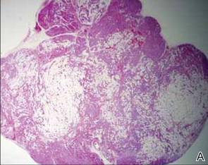
    A fibrous papule is a common benign lesion that usually presents in adults on the face, especially on the lower portion of the nose.