From the Department of Dermatology, Kurume University School of Medicine, Japan.
The author reports no conflict of interest.
Correspondence: Chika Ohata, MD, PhD, Department of Dermatology, Kurume University School of Medicine, 67 Asahimachi, Kurume, Fukuoka, Japan 830-0011 (bboohay02@ybb.ne.jp).
Hailey-Hailey disease typically presents as suprabasal blisters with a perivascular and interstitial lymphocytic infiltrate. The differential diagnosis of Darier disease, herpesvirus infection, pemphigus foliaceus, and pemphigus vulgaris also is discussed.
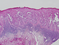
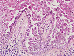
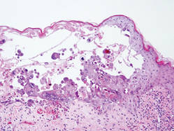
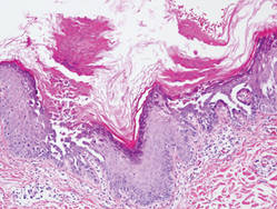
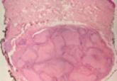
Hailey-Hailey disease (HHD), or benign familial chronic pemphigus, typically presents as suprabasal blisters with a perivascular and interstitial lymphocytic infiltrate (Figure 1).1 Villi, or elongated dermal papillae lined with a single layer of basal cells, protrude into the bullae (Figure 2). In HHD lesions, the epidermis is thickened with scale-crust, and at least the lower half of the epidermis shows acantholysis. Despite the acantholytic changes, a few intact intercellular bridges remain, giving the appearance of a dilapidated brick wall (Figure 2). There may be dyskeratotic cells among the acantholytic cells, though they are scant in many cases. These acantholytic dyskeratotic cells have eosinophilic polygonal-shaped cytoplasm. Hailey-Hailey disease typically does not show adnexal extension of the acantholysis. Direct immunofluorescence is negative in HHD.
Figure 1. A suprabasal blister with acantholytic changes in the lower half of the epidermis in the setting of Hailey-Hailey disease. A dense perivascular and interstitial lymphocytic infiltrate can be seen in the upper dermis (H&E, original magnification ×40).
Figure 2. Villi, or protruding dermal papillae lined with a single layer of basal cells, are evident. Above the villi, a few intact intercellular bridges remain, giving the appearance of a dilapidated brick wall (H&E, original magnification ×200).
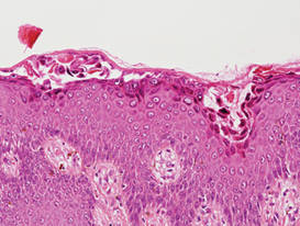
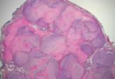
Pemphigus vulgaris is an autoimmune intraepidermal bullous disease that presents with suprabasal acantholysis (Figure 3).2 The epidermis is not thickened and acantholysis is limited to the suprabasal layer. Acantholytic cells with eosinophils and/or neutrophils are found within the bullae. Perivascular and interstitial infiltrates of lymphocytes, eosinophils, and occasionally neutrophils are seen; however, the inflammatory cell infiltrate can vary from extensive to scant. Direct immunofluorescence usually reveals IgG and/or C3 deposition on the surface of the keratinocytes throughout the epidermis.
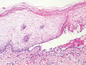
Pemphigus foliaceus is another autoimmune intraepidermal bullous disease that is characterized by acantholysis in the granular or upper spinous layers (Figure 4).3 The epidermis is not thickened. Sometimes acantholytic cells show dyskeratotic change (Figure 4). Some biopsy specimens do not contain the roof of the bullae; therefore, only erosion is seen and the diagnosis may be missed. Moreover, when only the adnexal epithelium shows acantholysis without epidermal involvement, diagnosis can be difficult.4 Acantholysis is accompanied with a superficial perivascular and interstitial inflammatory cell infiltrate consisting of lymphocytes, eosinophils, and occasionally neutrophils. The amount of inflammatory cell infiltrate may vary. Bullous impetigo and staphylococcal scalded skin syndrome reveal a similar histopathologic pattern. Direct immunofluorescence usually discloses IgG and/or C3 deposition on cell surfaces of keratinocytes in the entire or upper epidermis.
Figure 3. Intraepidermal bulla in pemphigus vulgaris caused by suprabasal acantholysis. A mixed infiltrate of lymphocytes and eosinophils is seen in the upper dermis (H&E, original magnification ×100).
Figure 4. Subcorneal acantholytic cells are evident. Some acantholytic cells are dyskeratotic in pemphigus foliaceus (H&E, original magnification ×200).
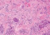
Herpesvirus infection shows ballooning (intracellular edema) of keratinocytes. Eventually acantholysis occurs and intraepidermal bullae are formed. In the bullae, virus-associated acantholytic keratinocytes, some that are multinucleated, can be easily found (Figure 5).5 These cells are larger than normal keratinocytes and have steel gray nuclei with peripheral accentuation. Some of these cells are necrotic, and the remains of necrotic multinucleated acantholytic cells are easily recognized. Adnexal epithelial cells occasionally are affected by herpesvirus infection; nuclear change is similar to the epidermis. A perivascular and interstitial infiltrate of lymphocytes and neutrophils is seen. Neutrophils accumulate within the old bullae, clinically manifesting as a pustule.
Darier disease is characterized by suprabasal clefts and acantholysis above the basal layer (Figure 6).6 Similar to HHD, villi protrude within the clefts (Figure 6). Conspicuous columns of parakeratosis above the acantholytic epidermis often are observed. Dyskeratotic cells exist among acantholytic ke-ratinocytes in the granular layer and parakeratotic column, which are known as corps ronds and crops grains, respectively. A scant to moderate lymphocytic infiltrate is found in the upper dermis.
Figure 5. Multinucleated cells with steel gray nuclei are easily found in a blister caused by herpesvirus infection (H&E, original magnification ×100).
Figure 6. Narrow foci of suprabasal clefts are seen intermittently in Darier disease. Above the suprabasal clefts, acantholytic changes with occasional acantholytic dyskeratotic cells throughout the epidermis are seen with columns of parakeratosis. Villi also are seen, similar to Hailey-Hailey disease (H&E, original magnification ×100).