Case Letter
Pedunculated Scrotal Nodule in an Elderly Male: A Rare Presentation of Trichoblastoma
Trichoblastoma is a rare benign tumor with differentiation toward the primitive hair follicle. Trichoblastomas show no gender predilection and...
From the Department of Pathology and Microbiology, University of Nebraska Medical Center, Omaha.
The authors report no conflict of interest.
Correspondence: Dominick J. DiMaio, MD, Department of Pathology and Microbiology, 983135 Nebraska Medical Center, Omaha, NE 68198-3135 (ddimaio@unmc.edu).

Trichoblastomas are rare cutaneous tumors arising from the hair bulb and mesenchyme. Although they are benign, they can pose a diagnostic dilemma for the clinician and pathologist because they clinically and histologically mimic more common lesions such as basal cell carcinomas (BCCs) and trichoepitheliomas. It is important for the clinician and pathologist to be aware of such tumors and their variants. We present a case of a melanotrichoblastoma, an exceedingly rare variant of trichoblastoma, as well as review the current literature on the clinical presentation and histologic differentiation of these unique tumors with their more commonly seen mimics.
Practice Points
Trichoblastomas are rare cutaneous tumors that recapitulate the germinative hair bulb and the surrounding mesenchyme. Although benign, they can present diagnostic difficulties for both the clinician and pathologist because of their rarity and overlap both clinically and microscopically with other follicular neoplasms as well as basal cell carcinoma (BCC). Several classification schemes for hair follicle neoplasms have been established based on the relative proportions of epithelial and mesenchymal components as well as stromal inductive change, but nomenclature continues to be problematic, as individual neoplasms show varying degrees of differentiation that do not always uniformly fit within these categories.1,2 One of these established categories is a pigmented trichoblastoma.3 An exceedingly rare variant of a pigmented trichoblastoma referred to as melanotrichoblastoma was first described in 20024 and has only been documented in 3 cases, according to a PubMed search of articles indexed for MEDLINE using the term melanotrichoblastoma.4-6 We report another case of this rare tumor and review the literature on this unique group of tumors.
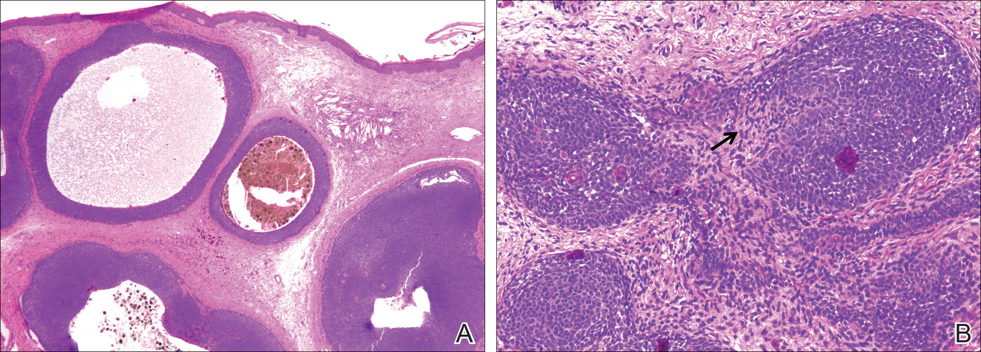
A 25-year-old white woman with a medical history of chronic migraines, myofascial syndrome, and Arnold-Chiari malformation type I presented to dermatology with a 1.5-cm, pedunculated, well-circumscribed tumor on the left side of the scalp (Figure 1). The tumor was grossly flesh colored with heterogeneous areas of dark pigmentation. Microscopic examination demonstrated that within the superficial and deep dermis were variable-sized nests of basaloid cells. Some of the nests had large central cystic spaces with brown pigment within some of these spaces and focal pigmentation of the basaloid cells (Figure 2A). Focal areas of keratinization were present. Mitotic figures were easily identified; however, no atypical mitotic figures were present. Areas of peripheral palisading were present but there was no retraction artifact. Connection to the overlying epidermis was not identified. Surrounding the basaloid nodules was a mildly cellular proliferation of cytologically bland spindle cells. Occasional pigment-laden macrophages were present in the dermis. Focal areas suggestive of papillary mesenchymal body formation were present (Figure 2B). Immunohistochemical staining for Melan-A was performed and demonstrated the presence of a prominent number of melanocytes in some of the nests (Figure 3) and minimal to no melanocytes in other nests. There was no evidence of a melanocytic lesion involving the overlying epidermis. Features of nevus sebaceus were not present. Immunohistochemical staining for cytokeratin (CK) 20 was performed and demonstrated no notable number of Merkel cells within the lesion.

Figure 2. Melanotrichoblastoma histopathology with variable-sized nests of basaloid cells with central cystic spaces. Pigmentation is associated with some of the nests (A)(H&E, original magnification ×20). A nest of basaloid cells on the right (black arrow) demonstrated an area of indentation in which there was peripheral palisading and condensation of stromal cells consistent with papillary mesenchymal body formation (B)(H&E, original magnification ×100).
Trichoblastoma is a rare benign tumor with differentiation toward the primitive hair follicle. Trichoblastomas show no gender predilection and...

An association between steatocystoma multiplex (SCM) and eruptive vellus hair cysts (EVHCs) has been recognized. Steatocystoma multiplex and EVHC...
The occurrence of cylindromas, trichoepitheliomas, and spiradenomas completes the triad for Brooke-Spiegler syndrome (BSS). This combination...
