Dr. Yari was from and Drs. Malone and Rivas are from the University of Texas Medical Branch, Galveston. Dr. Yari was from the Department of Internal Medicine, Dr. Malone is from the Department of Dermatology, and Dr. Rivas is from the Department of Internal Medicine. Dr. Yari currently is from the Department of Neurology, Baylor Scott & White Health, Texas A&M University Health Center, Temple.
The authors report no conflict of interest.
Correspondence: Niloofar Yari, MD, Department of Neurology, Baylor Scott & White Health, 2401 S 31st, MS-01-E524, Temple, TX 76508 (niloofar.yari@bswhealth.org).
Crusted scabies is a severe, highly contagious form of classic scabies caused by the mite Sarcoptes scabiei var hominis.Crusted scabies is more common in immunosuppressed populations and overcrowded environments. In this condition, the host’s immune system is overwhelmed and unable to defend against the mites on the skin, resulting in hyperinfestation of the host. Diagnosis can be challenging because the condition resembles other common skin conditions, such as plaque psoriasis. Furthermore, delayed diagnosis and inappropriate treatment can lead to worsening of the condition. We report a case of crusted scabies that was initially misdiagnosed in a 34-year-old incarcerated man with multidrug-resistant human immunodeficiency virus/AIDS. The patient had a complicated but complete recovery after treatment with permethrin and ivermectin was instituted.
Keep scabies in mind, especially in immunocompromised patients or populations in overcrowded areas.
Treatment consists of isolating the patient, starting topical permethrin and oral ivermectin (in severe cases), washing all linens, and prophylactically treating contacts.
References
Case Report
A recently incarcerated34-year-old man with an 11-year history of multidrug-resistant human immunodeficiency virus/AIDS (CD4 count, 121 cells/mm3; viral load, 49,625 particles/mm3 one week prior to presentation) was admitted to the hospital for an intensely pruritic, hyperkeratotic, scaly rash involving the entire body. The rash first appeared on the feet approximately 1 year prior to admission. At that time the patient was given oral fluconazole and a steroid cream with near resolution of the rash. He was then transferred multiple times to different units with subsequent discontinuation of the medications. The rash flared and progressed to involve the knees. He was restarted on the fluconazole and steroid cream and placed in isolation by medical personnel at the prison 6 months prior to presentation. The rash continued to spread, and he was given a working diagnosis of plaque-type psoriasis by several providers after several months of nonresponse to treatment. Additional attempts at treatment at outside facilities included oral fluconazole, trimethoprim-sulfamethoxazole, and other antibiotics. He was referred to dermatology at our institution but missed the appointment and was admitted to the hospital before the appointment could be rescheduled.
On admission to the hospital, he denied similar lesions in close contacts. On review of systems he had subjective fevers and chills, decreased appetite, nausea without vomiting, dysphagia to solids, epigastric pain, and 70-lb weight loss over the last 6 months. Facial involvement of the rash impaired the ability to open the mouth, speak, and eat. He had no known drug allergies. His only medications at the time of admission were nortriptyline, trimethoprim-sulfamethoxazole, and oral combination elvitegravir-cobicistat-emtricitabine-tenofovir for hu-man immunodeficiency virus treatment.
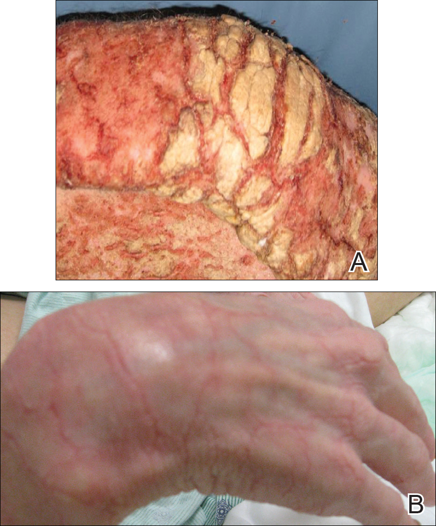
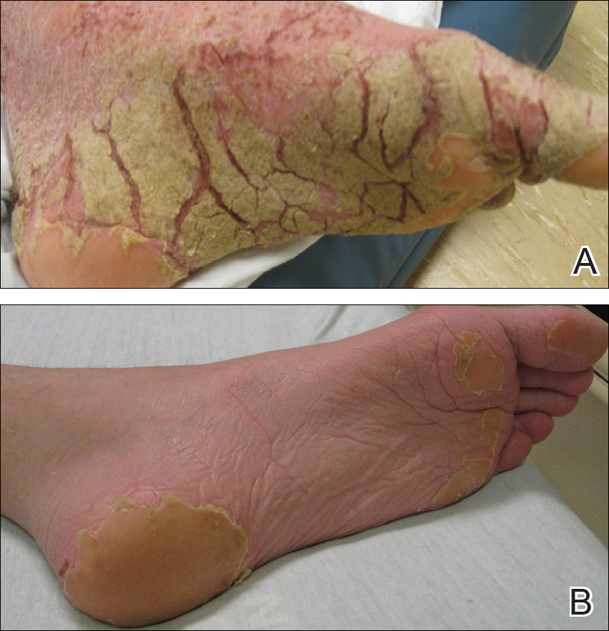
On physical examination he was cachectic, shivering, and foul smelling. He was afebrile, slightly tachycardic (112 beats per minute), and hypertensive (144/83 mm Hg) with a respiratory rate of 18 breaths per minute. His height was 1.83 m (6 ft) and weight was 48.5 kg (107 lb) with a body mass index of 14.5. Extensive erythematous, hyperkeratotic, crusted, and fissured plaques covered the entire body including the face, hands, and feet. The tongue was covered with bilateral white-colored plaques, and he had patches of alopecia, excoriations, and scales on the scalp. The elbows were fixed in a flexed position and he had decreased range of motion in the wrists and fingers due to the severe hyperkeratosis (Figure 1A). Hyperkeratosis also was prominent on the knees and feet with associated burrows (Figure 2A). He had foot drop on the left.
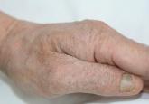
Figure 1. Hyperkeratotic lesions on the right hand before (A) and after 3 weeks of treatment with permethrin cream 5% and oral ivermectin (B).
Figure 2. Hyperkeratosis and visible burrows on the left foot before (A) and after 3 weeks of treatment with permethrin cream 5% and oral ivermectin (B).
The differential diagnosis included a drug eruption; fungal or parasite infestation, such as crusted scabies; psoriasis; or cutaneous lymphoma. Laboratory studies were difficult to obtain, as there were limited areas suitable for vascular access. Blood work showed leukocytosis (18.9×10⁹ cells/L [reference range, 4.8–10.8×10⁹ cells/L) with 13.3% eosinophils (reference range, 1%–6%). This eosinophilia narrowed the likely diagnoses to a drug eruption or parasite infection.
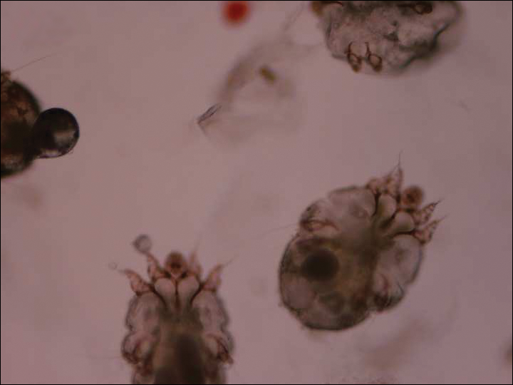
The dermatology service was consulted. A mineral oil preparation was performed and showed numerous mites and feces consistent with a diagnosis of crusted scabies (Figure 3). The patient was started on a regimen of permethrin cream 5% applied to the entire body, except the face, which was left on overnight and washed off. This regimen was repeated daily for 1 week, then twice weekly until the rash resolved after a total of 3 weeks. Due to the severity of his condition, immunocompromised status, and concern for superinfection, oral ivermectin 200 μg/kg once daily was added on days 1, 2, 8, 9, 15, 22, and 29.1
Figure 3. Mineral oil preparation showed scabies mites. The diagnosis was made after the mites were visualized under the microscope (original magnification ×400). Photograph provided by Matthew Petit, DO (Galveston, Texas).
Our patient’s hospital course was further complicated by symptomatic hypoglycemia, altered mental status, and superimposed methicillin-resistant Staphylococcus aureus bacteremia, as well as Pseudomonas aeruginosa bacteremia, pneumonia, and coffee ground emesis. He was transferred to the intensive care unit but fortunately did not require intubation. His overall condition, mental status, and rash gradually improved. Three weeks after admission he only had a few residual lesions on the feet with clearing elsewhere (Figures 1B and 2B). He was discharged with a skin moisturizer and was referred for physical and occupational therapy. On follow-up clinic visits at 3 and 6 months, he had recovered well with general improvement in his condition.