DermaDiagnosis
Man Is in Thick of Skin Problem
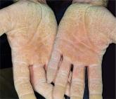
Since birth, a now 60-year-old man has been affected by the same dermatologic condition as several members of his mother’s family. As he has aged...

Ear “Wart” Prompts Unkind Comments
An 8-year-old boy is referred to dermatology for evaluation and treatment of a “wart” on the inferior rim of his left helix that has been present (and unchanged) since birth. The lesion is asymptomatic, and the boy’s biggest complaint is that it makes him the object of unkind comments from his siblings and friends. The patient’s mother claims the child is otherwise healthy; there is no history of seizure or other neurologic problems, and he does not have any medical conditions requiring treatment. The lesion has been treated, unsuccessfully, with a variety of wart remedies, including salicylic acid-based products and liquid nitrogen. Along the inferior rim of the left helix is a 5-cm linear collection of soft, skin-colored papules that range in size from pinpoint to 2.5 mm. They are so small and flesh-toned as to easily escape detection unless specifically sought. No other significant lesions are seen on the ear or elsewhere on the head or neck. The child looks his stated age and appears well developed and well nourished.
Since birth, a now 60-year-old man has been affected by the same dermatologic condition as several members of his mother’s family. As he has aged...
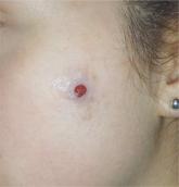
The lesion on the face of this 16-year-old girl is slightly tender to the touch and bleeds copiously with even minor trauma.
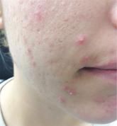
An 18-year-old woman is brought in by her mother for evaluation of longstanding acne.