
Radiology Review
From Hydroplane to Ankle Pain
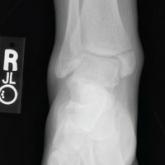
Following a car accident, a 40-year-old woman finds that bearing weight on her right foot causes excruciating pain. See if a radiograph can...

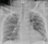
A 60-year-old man presents to the emergency department for evaluation of chest pain that began a few hours ago. He denies injury and has no associated nausea or shortness of breath. Earlier today, he underwent biopsy of a recently discovered mass in his right lung. Otherwise, his medical history is only significant for hypertension. He is a former pack-a-day smoker but quit three months ago.
On physical exam, you note an uncomfortable male in no obvious distress. He is afebrile, with normal vital signs. His O2 saturation is 96% on room air. Breath sounds appear to be clear bilaterally, although the patient expresses some discomfort with inhalation. Heart sounds are normal as well.
While the nurse and tech place an IV, a portable chest radiograph is obtained. What is your impression?
Following a car accident, a 40-year-old woman finds that bearing weight on her right foot causes excruciating pain. See if a radiograph can...
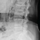
An 80-year-old woman with severe dementia chronically complains about her "ailments." But when her complaints of back pain intensify, a radiograph...
Following a motorcycle accident, a 55-year-old man complains of head, face, and chest wall pain. A chest radiograph clarifies some of the damage...