Family members bring in an 80-year-old man for urgent evaluation of a number of skin lesions. Most alarming to them is the lesion on his right ear, although many others, including numerous dark lesions on his back, have also raised their concern. The patient and his wife are “positive” all the lesions have been present, unchanged, for years.
EXAMINATION
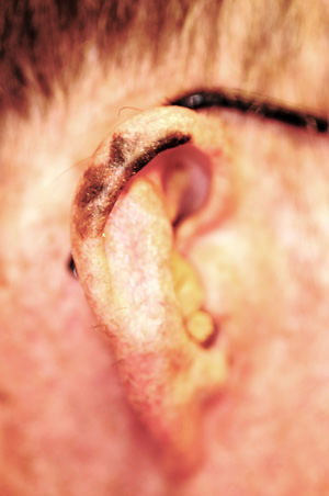
Your attention is quickly drawn to the superior helical area of the patient’s right ear, where a large, irregularly pigmented and bordered black patch covers most of the superior helix. The surrounding skin is quite fair and sun-damaged, with extensive solar elastosis seen all across the patient’s face—especially on the forehead, where multiple actinic keratoses are also seen and felt.
Hundreds of dark brown–to-black warty epidermal papules, nodules, and plaques are observed on the patient’s trunk; some are as large as 6 cm, while most average 2 to 3 cm. Fortunately, no other worrisome lesions are seen, except for the right superior helical lesion, which appears exceptionally suspicious for melanoma.
A tray is set up for the performance of a 3-mm punch biopsy of the ear lesion. Prior to that procedure, direct examination of the lesion is carried out with a dermatoscope, a magnifying (10x power) handheld viewing device that illuminates the site with flat, polarized light. It can be used to look for specific features of benign versus malignant lesions. After the entire lesion’s surface has been examined, the biopsy is cancelled and the family reassured regarding the benignancy of the ear lesion.
DISCUSSION
One reason for the dermatoscopic examination was the size of the ear lesion; it was so large that the chance of sampling error became a definite concern. A single 3-mm punch biopsy taken from a 4-cm lesion, if negative for cancer, could simply have missed the malignancy. However, removal of the entire lesion was not at all practical. Multiple punch biopsies from several areas within the lesion would have been an acceptable, but clumsy, alternative.
Thankfully, the dermatoscopic examination results obviated the need for invasive alternatives. Here’s why: Seen with the dermatoscope, over the entire surface of the lesion, were white pinpoint spots at regular intervals, called pseudocysts. These indentations are filled with keratin and are pathognomic for the diagnosis of seborrheic keratosis (SK). Moreover, no organized collections of pigment (streaks, globules, or networks) were seen; these might have suggested melanoma. The presence of so many other SKs elsewhere lent credence to that same diagnosis on the ear.
Even though SKs are often seen in non–sun-exposed areas, there is evidence to suggest that ultraviolet exposure can play a part in their genesis. Heredity and age are arguably more significant factors; SKs are seldom seen before the fourth decade of life.
SKs have little if any malignant potential, but they have been known to coincide with sun-caused skin cancers, such as basal or squamous cell carcinoma and melanoma, with one adjacent to or even overlying the other. And while the lesions of SK are typically raised and warty (epidermal or “stuck on”), they can appear quite flat and smooth at times, in addition to occasionally being darker than SKs are “supposed to be,” increasing their resemblance to a melanoma.
Biopsy of an SK shows diagnostic features of variable papillomatosis, as well as comedone-like openings, fissures, and keratin-filled pseudocysts. The differential for SKs includes wart, melanoma, and solar lentigo.
No treatment was attempted for this helical SK. However, the patient will be monitored during twice-yearly follow-up visits to dermatology, not only for changes to his ear lesion but also for changes anywhere else on his skin.
LEARNING POINTS
• Seborrheic keratosis (SK) is the most common example of a benign epidermal (“stuck-on”) lesion.
• That “stuck-on” nature is what usually distinguishes SK from melanoma.
• However, some SKs are almost completely flat and occasionally totally black—and therefore difficult to distinguish from melanoma, especially in sebum-rich skin.
• Biopsy is often necessary, but dermatoscopic examination (in trained hands) is a useful noninvasive procedure that can obviate the need for biopsy.