An 84-year-old woman has had a lesion on her face for 10 years. During that time, it has slowly changed, becoming darker and larger. For the past few months, it has developed a new, raised area in its center.
Several providers, including a dermatologist, have seen the lesion over the years; none has ever ordered a biopsy. The lesion is asymptomatic and only began to change noticeably a few years ago.
The patient mentions having multiple sunburns as a young woman. But she says she never tried to tan, because she could only do so with difficulty.
Her medical history is significant for breast cancer, for which she is receiving chemotherapy. She was told she did not need surgery and has little chance of metastasis.
EXAMINATION
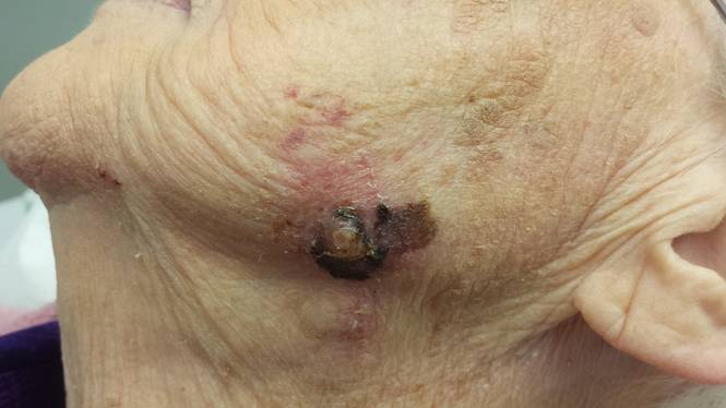
The patient is alert and cooperative. A large, black, 3.5-cm lesion is obvious on the right jawline. In the center of the lesion is a 2-cm, faintly eroded nodule. No tenderness can be elicited on palpation of the lesion or the area, and there are no palable nodes in the adjacent head and neck. The patient has type 2 skin, blue eyes, and modest evidence of sun damage elsewhere on her face.
DISCUSSION
Of course, the next logical step was to biopsy the lesion. The rule of thumb for biopsy of a possible melanoma is this: Removal of the entire lesion, down to adipose tissue, is the gold standard, because it is certain to provide both an accurate diagnosis and an assessment of the lesion’s vertical thickness—a key predictor of prognosis.
The drawback to complete excision of lesions this size is that the defect can be huge (about 10 cm in this case) and almost certainly disfiguring. If the lesion turns out to be benign, this outcome might well be seen as unacceptable. Therefore, an alternative to complete excision is incisional biopsy, in which a significant but modest portion of the lesion is removed, purposely obtaining the darkest, most irregular part. In this case, such a biopsy would include either all or a significant part of the nodular portion of the lesion, along with a generous section of the discolored macular area.
Shave biopsy of a suspected melanoma is acceptable with small, shallow lesions, as long as it effectively removes the lesion with deep margins. Shave biopsy in this case would be unacceptable for several reasons. Among them is the cosmetic outcome and the possibility of transecting the lesion, thereby making an accurate vertical assessment difficult. The latter is important, since it is the single most accurate prognostic indicator, in effect dictating the margins of re-excision as well as the extent of subsequent workup (imaging, bloodwork, lymph node dissection, and possible chemotherapeutic regimen).
Punch biopsy of suspected melanoma is generally unacceptable, unless the punch effectively removes most or all of the lesion. Referral to dermatology is almost never the wrong thing to do, but it often requires provider-to-provider communication to expedite.
Providers who don’t perform these biopsies often tend to quote the misinformed attending physicians who taught them that “cutting through a melanoma will make it spread.” This is a totally unfounded fear, long since disproven in large studies that showed no difference in mortality or morbidity between patients whose melanomas were incised versus those whose cancers were removed without incision.
DIAGNOSIS AND TREATMENT
Incisional biopsy was performed on this lesion, with about one-third removed by wedge incision. Essentially, the center of the lesion was removed. This was, of course, done under local and sterile conditions, with closure by simple interrupted sutures.
Within 48 hours, the report was issued. It showed a nodular melanoma with 3.7-mm Breslow thickness, definite signs of focal ulceration, and an increased mitotic rate of 6 to 8 per high-powered field—all of which predict a less-than-favorable prognosis.
The patient was scheduled for Mohs surgery, which will likely involve wide excision of the lesion and surrounding skin. She will also have the option of lymph node dissection, done for prognostic value only (as it will add nothing to her potential for survival).
TAKE-HOME LEARNING POINTS
• Biopsy of melanoma (or other skin cancers) cannot cause it to spread.
• The object of biopsying a potential melanoma is to obtain a specimen that, on pathologic examination, not only conveys the correct diagnosis but also allows for accurate staging.
• Successful biopsy entails going deep and wide enough, but without causing unnecessary scarring or deformation.
• The diagnosis and staging then determine the extent of any re-excision and the need for ancillary studies (eg, lymph node dissection, imaging, referral to oncology).