DermaDiagnosis

Two Different Diagnoses, But One Location

Author and Disclosure Information


A 57-year-old man self-refers for evaluation of a facial lesion that has been present for several years. As the lesion slowly grew larger, family members became worried and urged him to seek treatment. The man has spent many years in the sun during his adult life, both at work and when engaging in various hobbies, and he burns easily and often. In addition, 20 years ago, he was diagnosed with psoriasis, which was eventually treated with a year’s worth of phototherapy. History also includes a coronary artery bypass graft (undergone 10 years ago) and current smoker status. On examination, the facial lesion proves to be macular, with ominously irregular colors of black and brown, along with ragged borders. The overall size is almost 4 × 2 cm. The lesion is seen in the context of very fair, sun-damaged skin, characterized by widespread actinic keratoses and telangiectasias. Through use of a dermatoscope (a power-viewing instrument that provides 10× magnification, used to examine suspicious lesions for specific changes suggestive of cancer), definite signs of seborrheic keratosis (SK; pseudocysts) are seen. Nonetheless, the overall appearance of the lesion—as well as the patient’s high-risk status—dictates that a punch biopsy be performed. The subsequent report reveals the lesion to be a Clark’s level II superficial melanoma, Breslow 0.6 mm.

The dermatoscopic findings and biopsy results indicate two different diagnoses because:

a) The dermatoscopic findings were misinterpreted.

b) Melanoma can display pseudocystic features.

c) SK and melanoma can coincide in the same location.

d) The biopsy report was wrong.

ANSWER
The white, round spots seen in this lesion were entirely consistent with SK and were not misinterpreted (choice “a”). Since melanoma is not known to display this type of spot, choice “b” is also incorrect. Biopsy reports are occasionally wrong, of course, but the diagnosis of melanoma is fraught with such serious implications that the call is not made if specific criteria are not met. These include specific stains for that diagnosis, histologic findings, and the concurrence of multiple observers, so it is unlikely that the histologic diagnosis is incorrect (choice “d”).
This leaves the correct answer, choice “c,” discussed below.

DISCUSSION
There is truly no law saying that two lesions can’t appear in the same location—and as this case illustrates, it definitely happens. Had we performed additional biopsies, no doubt features of SK would have been seen in corroboration of the dermatoscopic findings.

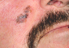
This case also emphasizes another useful principle: Pay just as much attention to the owner as to the lesion itself. This patient was quite fair and sun-damaged, so any odd lesion he developed would be suspect, let alone one that looked like this.
One final thing to be learned from this case is that as useful as dermatoscopes can be, they still must take a backseat to common sense.

Next Article: