BY CATALINA MATIZ, MD, AND ANDREA WALDMAN, MD
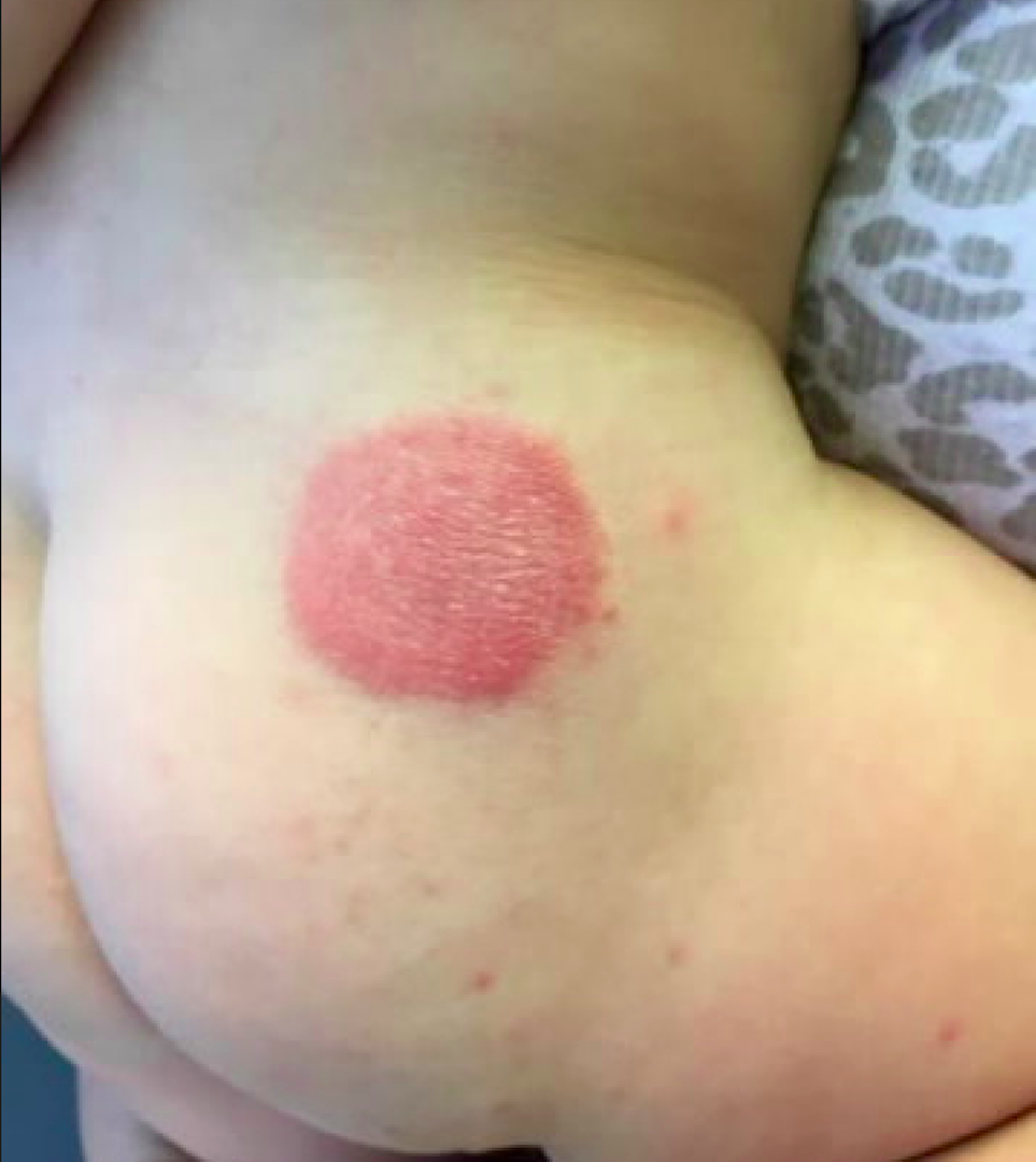
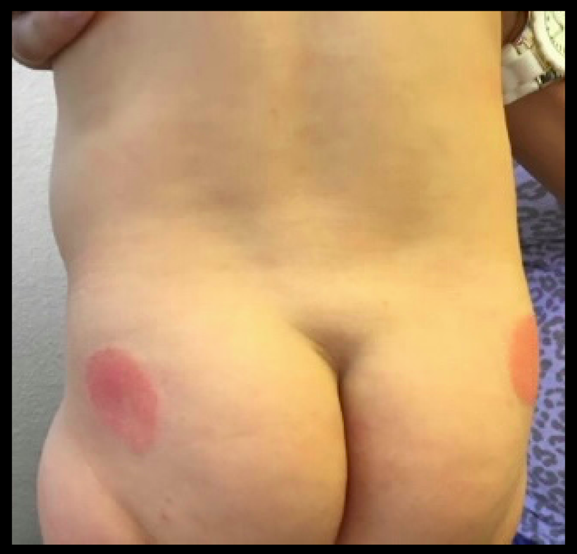
At the time of initial evaluation, the patient was prescribed a 2-week course of topical triamcinolone 0.1% ointment. Subsequently, the cutaneous lesions clinically improved, but did not completely resolve. Mother had no recollection of infant contact with metal, plastic, or additional topical emollients or medications. On further investigation, the patient’s mother revealed the recent conversion to a different brand of diaper. The diapers had a blue and green polka dot design corresponding with the distribution of our patient’s cutaneous lesions, highly suggestive of allergic contact dermatitis (ACD).
Diaper dermatitis represents a common complaint peaking at 9-12 months of age, estimated to occur in 25% of infants in the primary care office setting.1 Irritant contact dermatitis (ICD) predominates as the most common etiology of diaper dermatitis. ICD represents a nonimmunologic response, triggered by maceration and breakdown of the cutaneous barrier, directly resulting from chronic exposure to urine and feces.1 In contrast, ACD characterizes an immunologic reaction to specific offending allergen(s).
ACD can arise in the diaper area following contact with dyes, fragrances, or other constituents of disposable diapers, wet wipes, diaper creams, or medications. Alberta et al. initially described ACD in several infants, resulting from blue, green, and pink dyes present in various disposable diapers. The dermatitis promptly resolved upon switching to dye-free diapers.2 Furthermore, topical products applied to the diaper region such as barrier creams and wet wipes may trigger allergic sensitization. Sensitization to an allergen can occur after few exposures to years after introduction. Following the initial contact reaction, re-exposure to the offending allergen can elicit a quicker cutaneous response. Case reports have detailed cases of ACD presenting in neonates as early as 1 week of age.3
If ACD is suspected as an etiologic factor in diaper dermatitis, providers should consider constituents present in topical formulations, wet wipes, and components of the diaper itself. A specific example includes mercaptobenzothiazole, a rubber additive commonly found in elastic bands of disposable diapers.2 The most common potential allergens discovered in a recent review of 63 diaper wipes, 41 topical diaper preparations, and 3 top-selling diaper brands included botanical extracts (i.e. aloe vera), alpha-tocopherol, fragrances, propylene glycol, parabens, iodopropynyl butylcarbamate, and lanolin.4
ACD may be difficult to diagnose. Obtaining a thorough clinical history is essential to diagnosis. ACD is often distinguished by its distinct cutaneous morphology, with initial lesions commonly characterized by sharply demarcated erythema associated with vesiculation and edema. The dermatitis progressively evolves to a mixed presentation of predominantly eczematous papules and plaques with crusting, scaling, and lichenification. Another clue suggestive of ACD is cutaneous involvement limited to areas of allergen contact, such as the geometric plaques corresponding to areas of skin exposure to blue and green dye in our patient.1
Differential diagnosis
In contrast to ACD, ICD commonly erupts in the perianal region in the presence of feces, especially during diarrheal episodes. Common clinical manifestations include skin maceration, erosions, erythema, papulation, and scaling. More severe presentations of irritant dermatitis include Jacquet’s diaper dermatitis, perianal pseudoverrucous papules and nodules and granuloma gluteale infantum.1 These cutaneous findings also may develop in older patients with irritated peristomal skin.5
Inflammatory dermatoses such as psoriasis and seborrheic dermatitis also may erupt in the diaper region. Psoriasis commonly involves the gluteal cleft and inguinal folds, appearing as well-demarcated, pink plaques with thin white scale.5 Persistence of plaques despite topical corticosteroid application and lack of pruritus further delineates this disorder. Family history of psoriasis may provide an additional clue to the diagnosis of psoriasis. Similarly, seborrheic dermatitis may present with diaper involvement characterized by well-demarcated erythematous plaques with minimal or absent greasy yellow scale. This dermatitis commonly erupts in infants and neonates less than 6 weeks old and resolves by 6-9 months of age.1
Langerhans cell histiocytosis (LCH) represents a serious condition important to consider in the differential diagnosis of persistent recalcitrant diaper dermatitis. Often confused with seborrheic dermatitis, the diaper dermatitis of LCH may be differentiated by petechiae, purpura, and/or ulceration and atrophy of the inguinal folds.5
Management
Avoidance of the allergen constitutes the gold standard of treatment for ACD. Initial treatment involves adequate-potency topical corticosteroids; for the diaper area, low- to mid-potency corticosteroids are recommended. Changing to dye-free diapers and/or avoiding contact with the suspected allergen also is advised. Epicutaneous patch testing is recommended if the dermatitis fails to improve despite removal of the suspected offending agent.5
References
1. Pediatr Dermatol. 2014 Nov;31 Suppl 1:19-24.
2. Pediatrics. 2005 Sep;116(3):e450-2.
3. Cutis. 1994 Nov;54(5):300-2.
4. Dermatitis. 2016 May-Jun;27(3):110-8.
5. “Neonatal and Infant Dermatology,” 3rd edition (Philadelphia: Elsevier, 2015, p. 251)
Dr. Matiz is assistant professor of dermatology at Rady Children’s Hospital San Diego–University of California, San Diego, and Dr. Waldman is a clinical research fellow at the hospital. Email them at pdnews@frontlinemedcom.com.