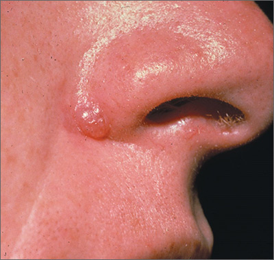
One week later, the pathologist reported that the growth was an amelanotic melanoma of 1.2 mm depth. The FP was relieved that he sent the tissue for pathology and did not assume this was a benign pyogenic granuloma. The patient was referred to a head and neck surgeon for complete excision with margins and a sentinel lymph node biopsy. She was fortunate to not have any nodal metastases. The FP performed a complete skin exam and found no other lesions suspicious for melanoma.
Photos and text for Photo Rounds Friday courtesy of Richard P. Usatine, MD. This case was adapted from: Smith M. Usatine R. Pyogenic Granuloma. In: Usatine R, Smith M, Mayeaux EJ, et al, eds. Color Atlas of Family Medicine . 2nd ed. New York, NY: McGraw-Hill; 2013: 940-944.
To learn more about the Color Atlas of Family Medicine , see: www.amazon.com/Color-Family-Medicine-Richard-Usatine/dp/0071769641/
You can now get the second edition of the Color Atlas of Family Medicine as an app by clicking on this link: usatinemedia.com