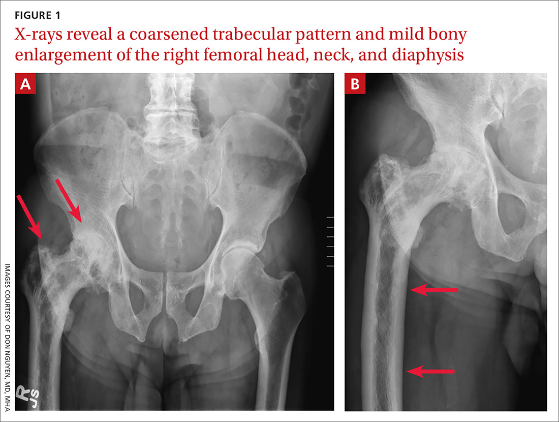
A 65-year-old man with a history of remote colon cancer, peptic ulcer disease, gastroesophageal reflux disease (GERD), and bilateral knee replacements presented with right groin and hip pain of more than a year’s duration. The patient described his hip pain as aching and said that it had worsened over the previous 6 months, interfering with his sleep. He said the pain worsened following activity, and it briefly felt better following an intra-articular corticosteroid injection into his right hip. The patient denied recent trauma or fracture and said he had no scalp pain, hearing loss, or spinal tenderness. Physical examination showed limited range of motion of the right hip and mild tenderness to palpation. Laboratory values were within normal limits. X-rays of the pelvis (Figure 1A) and right hip (Figure 1B) were ordered.
Photo Rounds
Right hip and pelvic pain
J Fam Pract. 2020 January;69(1):43-46
Author and Disclosure Information
Brigham and Women’s Hospital, Boston, MA (Dr. Nguyen); Medical University of South Carolina, Charleston (Dr. O’Connor); Allegheny Health Network, PA (Dr. Tran); Beth Israel Deaconess Medical Center, Boston (Dr. Qureshi)
dnguyen42@bwh.harvard.edu
DEPARTMENT EDITOR
Richard P. Usatine, MD
University of Texas Health at San Antonio
The authors reported no potential conflict of interest relevant to this article.

Follow-up imaging confirmed a clinical suspicion.

