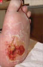
A 60-year-old woman came into our area hospital seeking relief for a foot wound that she’d had for several months (FIGURE 1A). The patient said that she had used several antibiotics (prescribed by her primary care physician over the past few months) and she changed her dressings daily, but the wound was not going away. Her podiatrist had evaluated her for osteomyelitis, and the results of the bone biopsy were pending.
The patient had a large wound on the plantar surface of her left foot. The skin over the surface had full-thickness breakdown of the epidermis and dermis, with partial necrosis of the subcutaneous tissue.
Following the wound edges did not reveal undermining, and there was no evidence of sinus tract formation. The ulcer did not extend through the fascia, and there was no gross damage to underlying muscle, bone, or tendon.
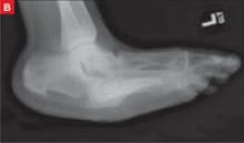
Her past medical history was significant for diabetes with neuropathy, nephropathy, and retinopathy. A radiograph was taken (FIGURE 1B).
FIGURE 1
Abnormally shaped foot and pressure ulcer
What is your diagnosis of her foot deformity?
What is the stage of her pressure ulcer?


