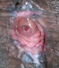
The FP recognized the white areas as leukoplakia suggestive of vulvar intraepithelial neoplasia (VIN). Punch biopsies showed moderate vulvar intraepithelial neoplasia II (VIN II) along with associated human papillomavirus (HPV) changes.
More than 50% of patients presenting with vulvar dysplasia have no symptoms. When symptoms are present, pruritus is the most common one. Unlike cervical intraepithelial neoplasia, VIN is usually multifocal. The most common locations include the interlabial grooves, posterior fourchette, and the perineum. Since VIN can appear warty, lesions that are diagnosed as condyloma (as was the case with this patient) and that don’t respond to conservative therapy should be biopsied to rule out VIN.
Biopsy to exclude invasive disease is mandatory prior to any topical treatment. Patients with VIN II or III should have their lesions removed. Basaloid and warty VINs may be treated with ablation on non–hair-bearing epithelium. A CO2 laser is typically used to remove the lesions, although some physicians perform a loop electrosurgical excision procedure.
Alternatives to surgery and laser include topical agents. 5-Fluorouracil cream, which causes a chemical desquamation of the lesion, has traditionally been used to treat VIN. It may result in significant burning, pain, inflammation, edema, and occasional painful ulcerations. Imiquimod cream is a topical immune response modifier that is FDA approved for the treatment of anogenital warts, actinic keratosis, and certain basal cell carcinomas. It has been used to treat multifocal VIN II or III in a few small pilot studies. The cream is self-administered 3 times per week for periods of 6 to 34 weeks.
In this case, the patient was referred for treatment of the VIN and given 2 g of oral metronidazole for the Trichomonas infection. She was advised to inform her most recent sexual partner about the Trichomonas infection so that he might receive treatment. Testing for other sexually transmitted diseases was negative.
Photo and text for Photo Rounds Friday courtesy of Richard P. Usatine, MD. This case was adapted from: Mayeaux EJ. Vulvar intraepithelial neoplasia. In: Usatine R, Smith M, Mayeaux EJ, et al, eds. Color Atlas of Family Medicine. 2nd ed. New York, NY: McGraw-Hill; 2013:519-524.
To learn more about the Color Atlas of Family Medicine, see: http://www.amazon.com/Color-Family-Medicine-Richard-Usatine/dp/0071769641/
You can now get the second edition of the Color Atlas of Family Medicine as an app by clicking on this link: http://usatinemedia.com/


