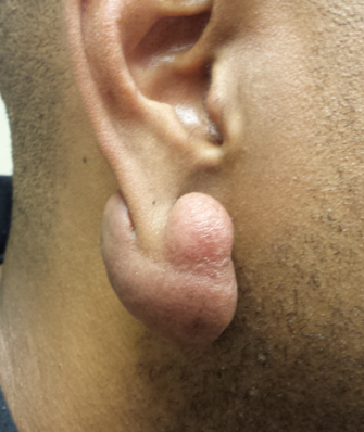
A 39-year-old African-American man has had a keloid on his right earlobe for several years. It was triggered by ear-piercing and has grown slowly to its current size. The patient denies any symptoms with the lesion.
EXAMINATION
The 5 x 2-cm lobulated mass is consistent in every way with a keloid: firm and pedunculated, with no epidermal changes. It hangs from his right earlobe. The patient has type IV skin, of African-American ancestry. After an extended discussion regarding treatment options, the patient decides to have the lesion excised.
PROCEDURE
Under sterile conditions and with local anesthesia (1% lidocaine with epinephrine), the entire keloid is removed. The wound edges are shaped to enhance the final appearance, and the wound is closed in one layer.
Since the risk for reformation is all too real, at one week post-op, the wound edges are injected with 3 mg/cc of triamcinolone, with a total of 1.5 cc used. The injections continue for three months as the wound heals. The outcome is totally acceptable to the patient, representing a huge improvement from his pre-operative appearance.
Continue reading for Joe Monroe's discussion...