DermaDiagnosis

Lesion Has Doubled in Size in Two Weeks

Author and Disclosure Information

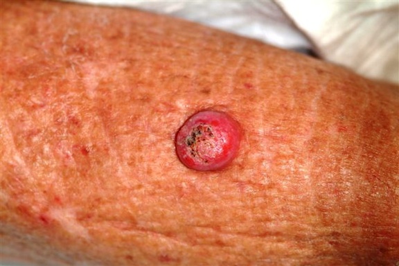
Woman with lesion on forearm.

The patient's lesion has grown rapidly, doubling in size in the past two weeks.

Six weeks ago, a new lesion began to appear on the forearm of this 63-year-old woman; it has grown rapidly in the ensuing time. She first went to her primary care provider, who diagnosed a staph infection and prescribed an antibiotic that hasn’t helped. The lesion, while asymptomatic, is nonetheless alarming; it has doubled in size in the past two weeks, which is why the patient now presents to the dermatology clinic. Her medical history includes well-controlled hypertension and a remote incidence of several basal cell carcinomas. On examination, the patient’s skin, in general, is severely sun-damaged, as evidenced by a weathered, leathery look to all exposed skin, as well as by multiple telangiectasias and solar lentigines. The lesion in question is a striking 1.8-cm dome-like nodule with a central keratotic core, located on the mid-dorsal forearm. Smooth and shiny, the surface of this firm nodule is also 
covered with tiny telangiectasias. Careful examination of the rest of the patient’s skin shows no other worrisome lesions.

Given the facts of the case, all of the following statements are probably true, except:

a) The lesion is almost certainly benign.

b) The lesion is probably a keratoacanthoma.

c) The lesion is at least in part caused by sun exposure.

d) Left alone, the lesion will likely resolve on its own.

ANSWER
The one incorrect statement is choice “a,” since the history and appearance of the lesion are quite consistent with keratoacanthoma—considered by most to be a low-grade form of squamous cell carcinoma (SCC). All the other statements are true.

DISCUSSION/TREATMENT
Keratoacanthomas (KAs) are quite common, especially in older patients with fair, sun-damaged skin. They appear on areas of the skin that have been directly exposed to the sun. The dorsal forearm is especially typical, but KAs can also appear on the face, hands, shoulders, and back.

The relatively rapid growth is in marked contrast to most other skin cancers and has been linked to the human papillomavirus DNA, which has been found in these lesions. However, by no means is this connection universally accepted as the cause, even though immune suppression, in the susceptible patient, does appear to play a role.

Microscopically, KAs bear a marked resemblance to SCCs. Indeed, they have been known to metastasize in rare instances, although most will involute (albeit with minor scarring) on their own, without treatment. The standard of treatment in this country is to remove KAs surgically and to submit the tissue for pathologic examination, which would officially show “SCC, KA type,” or “well-differentiated SCC.”

The differential diagnosis includes wart, invasive SCC, Merkel cell carcinoma, and melanoma. KAs have nothing to do with bacterial infection.

Next Article: